section name header

Basic Information ⬇

Author: Sajeev Handa, MD, SFHM and Michael Rossi, MD

Definition

Aspergillosis refers to several forms of a broad range of illnesses caused by infection with Aspergillus species. Aspergillus species is a ubiquitous mold most commonly found in soil, building materials, and water.

ICD-10CM CODES
B44.0Invasive pulmonary aspergillosis
B44.1Other pulmonary aspergillosis
B44.2Tonsillar aspergillosis
B44.7Disseminated aspergillosis
B44.81Allergic bronchopulmonary aspergillosis
B44.89Other forms of aspergillosis
B44.9Aspergillosis, unspecified
Epidemiology & Demographics
Incidence & Prevalence:

  • •Aspergillus species are ubiquitous in the environment internationally and occur as a mold found in soil. Aspergillus fumigatus is the most common human pathogen.1
  • •Aspergillus may cause a variety of illnesses from hypersensitivity pneumonitis to disseminated overwhelming infection in immunosuppressed patients.2
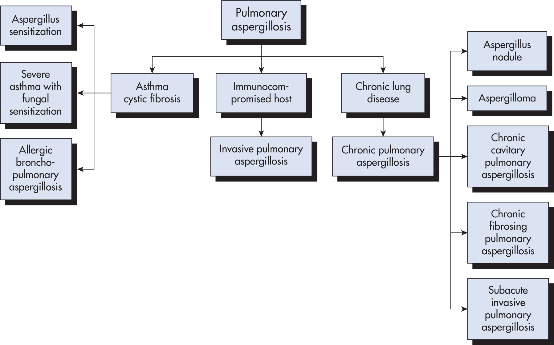
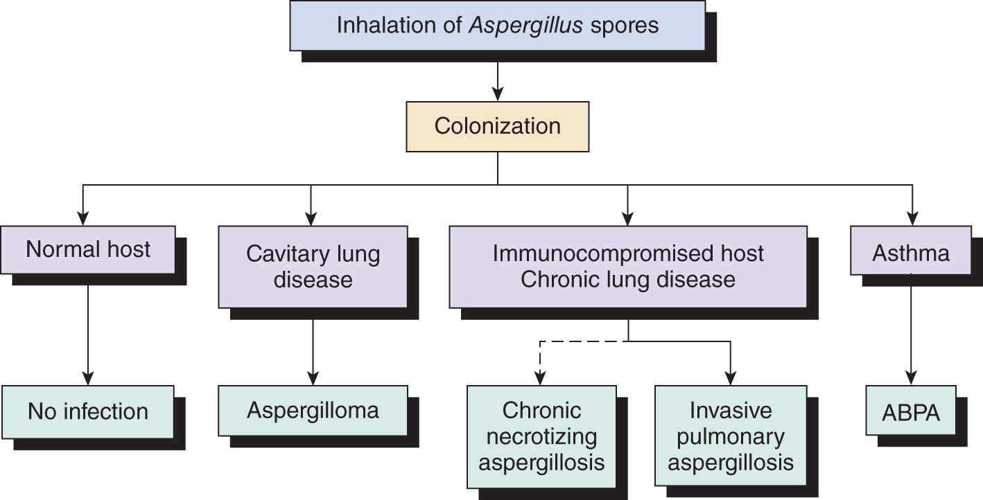
  • •Figs. E1 and E2 illustrate the categories of pulmonary aspergillosis.
  • •Frequently cultured from hospital wards from unfiltered outside air circulating through open windows as well as water sources.
  • •Reach the patient by airborne conidia (spores) that are small enough (2.5 to 3 μm) to reach the alveoli on inhalation.
  • •Can invade the nose, paranasal sinuses, external ear, or traumatized skin.

Figure E1 Categories of pulmonary aspergillosis.

!!flowchart!!

(From El-Baba F et al: Pulmonary aspergillosis: what the generalist needs to know, The American Journal of Medicine 133(6):668-674, 2020.)

Figure E2 The Spectrum of Aspergillus Infection

Abpa, Allergic Bronchopulmonary Aspergillosis.

!!flowchart!!

(From Sellke FW et al: Sabiston and Spencer: surgery of the chest, ed 10, Philadelphia, 2024, Elsevier.)

Risk Factors:

  • •The clinical syndrome depends on the underlying lung architecture, the host’s immune response, and the degree of inoculum.
  • •Incidence of invasive aspergillosis is increasing with advances in the treatment of life-threatening diseases, such as chemotherapy for hematologic malignancies or bone marrow and solid organ transplantation that are associated with suppression of the immune system and prolonged neutropenia. Allogeneic stem cell and lung transplant recipients are at highest risk for invasive pulmonary aspergillosis. Genetic deficiency of the soluble-pattern-recognition receptor known as long pentraxin 3 (PTX3) affects the antifungal capacity of neutrophils and may contribute to the risk of invasive aspergillosis in patients treated with hematopoietic stem-cell transplantation (HSCT).3
  • •Patients treated with the Bruton tyrosine kinase inhibitors (ibrutinib, acalabrutinib, and zanubrutinib) for chronic lymphocytic leukemia/small lymphocytic lymphoma are at risk for invasive aspergillosis.4
  • •Patients with AIDS and a CD4 count <50/mm3 have an increased susceptibility to invasive aspergillosis, but it is otherwise uncommon in patients with HIV.5
  • •Pandemic influenza A (H1N1) was initially associated with invasive pulmonary disease, and more recent studies have linked other influenza A and B strains with increased risk for invasive disease.
  • •COVID-19 secondary infection with invasive pulmonary Aspergillus infection has been seen in 2.5% to 35% of patients with severe acute respiratory distress syndrome (ARDS) requiring intubation and is associated with higher mortality.6
  • •Patients with chronic granulomatous disease are at higher risk for infections with Aspergillus species, and it is the most common cause of infectious death in chronic granulomatous disease (CGD).
  • •Aspergillus nidulans causes aggressive, invasive infections in patients with chronic granulomatous disease.
Etiology

  • •Aspergillus fumigatus is the usual cause.
  • •Aspergillus flavus is the second most important species, particularly in invasive disease of immunosuppressed patients and in lesions beginning in the nose and paranasal sinuses. Aspergillus niger can also cause invasive human infection.
Allergic Fungal Sinusitis:

  • •Occurs in patients with history of allergic rhinitis.
  • •Management involves aerating the sinus and ensuring tissue is not invaded via sinus CT.2
Allergic Bronchopulmonary Aspergillosis (Abpa):

  • •Symptoms occur most commonly in patients with asthma and cystic fibrosis.
  • •It is a long-term allergic response to Aspergillus.
  • •Results from an initial type I (immediate hypersensitivity) and type III reactions (immune complexes).
  • •Underdiagnosed pulmonary disorder in patients with asthma and cystic fibrosis and can present with recurrent episodes of asthma exacerbations. The reported prevalence in asthmatic patients varies from 1% to 2% and in cystic fibrosis from 2% to 15%.
Chronic Pulmonary Aspergillosis:

  • •A group of clinical syndromes related to aspergillus growth in underlying damaged lung tissue and includes: Aspergillomas ("Fungus Balls"), Aspergillus nodules, Chronic Cavitary, Chronic Fibrosing, and Subacute invasive Pulmonary Aspergillosis.
    1. 1.Diagnostic criteria for chronic pulmonary aspergillosis are summarized in Table E1.
    2. 2.In the absence of invasion or significant immune response, Aspergillus can colonize a preexisting cavity, causing a pulmonary aspergilloma.
  • •Aspergilloma involves formation of masses of hyphae in a prior pulmonary cavity.
  • •Patients typically have a history of chronic lung disease, e.g., tuberculosis, sarcoidosis, or emphysema.
  • •Manifests commonly as hemoptysis.
  • •Many are asymptomatic or minimally symptomatic for long periods of time.

TABLE E1 Diagnostic Criteria for Chronic Pulmonary Aspergillosis

  • •Thoracic radiologic findings consistent with the disease including one or more cavities with or without fungal ball or nodules (CT scan of the chest is preferred over plain films)
  • •Direct evidence of Aspergillus infection (microscopy or culture from biopsy) or an immunologic response to Aspergillus species (such as elevated serum Aspergillus IgG or precipitins, strongly positive serum or bronchoalveolar lavage galactomannan, or Aspergillus polymerase chain reaction)
  • •Exclusion of alternative diagnoses
  • •Duration of symptoms or radiologic changes for at least 3 mo

CT, Computed tomography; IgG, immunoglobulin G.

Reproduced with permission of the ERS 2025. Agarwal R, Sehgal IS, Muthu V, et al. Revised ISHAM-ABPA working group clinical practice guidelines for diagnosing, classifying and treating allergic bronchopulmonary aspergillosis/ mycoses. Eur Respir J 2024; 63: 2400061 [DOI: 10.1183/13993003.00061-2024]. This version is distributed under the terms of the Creative Commons Attribution Non-Commercial Licence 4.0.

Invasive Aspergillosis:

  • •In immunocompromised patients, presents as fever, focal pulmonary infiltrates, nodules, or wedge-shaped opacities.
  • •Typically a necrotizing bronchopneumonia, ranging from small areas of infiltrate to extensive bilateral hemorrhagic infarction.
  • •Most common presentation: Unremitting fever and a new pulmonary infiltrate despite broad-spectrum antibiotic therapy in an immunosuppressed patient.
  • •Dyspnea and nonproductive cough are common; sudden pleuritic pain and tachycardia, sometimes with a pleural rub, may mimic pulmonary embolism; hemoptysis may also be present.7
  • •Chest x-ray (CXR) may reveal patchy bronchopneumonic, nodular densities, consolidation, or cavitation. High-resolution CT scan (Fig. E3) is more sensitive and specific than CXR in neutropenic patients.
  • •Invasive aspergillosis may occur in the setting of severe influenza infections due to virus-induced respiratory epithelium even among immunocompetent hosts.8
  • •Immunocompromised patients: Invasive pulmonary Aspergillus (IPA) generally has an incubation period of around 15 days in neutropenic patients; invasive aspergillosis though, in general, is uncommon in the immunocompetent.2
  • •Criteria for classification of invasive pulmonary aspergillosis are summarized in Table E2.

Figure E3 Angioinvasive Aspergillosis

CT Section at the Level of the Lower Trachea Shows a Consolidation with an Eccentric Cavitation and Air Crescent Sign (Arrows). This Finding in This Neutropenic Patient is Highly Diagnostic of Angioinvasive Aspergillosis.
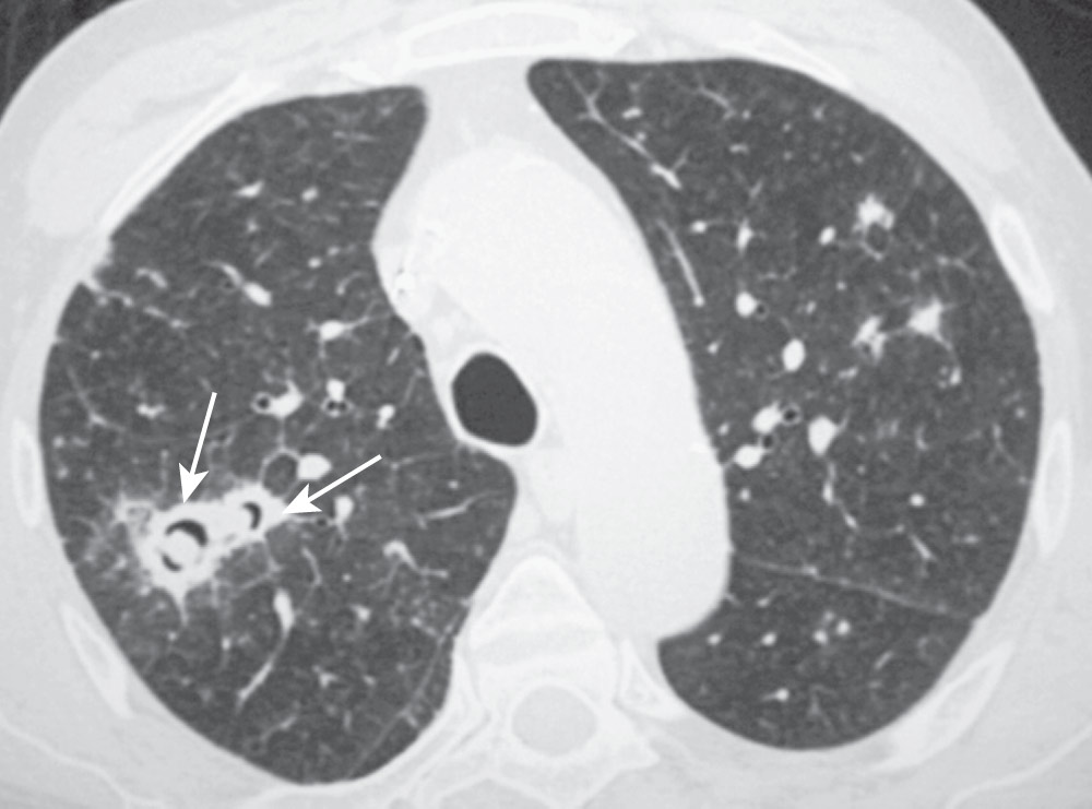
(From Franquet T: Nonneoplastic parenchymal lung disease. In Haaga JR, Boll DT [eds]: CT and MRI of the whole body, ed 6, Philadelphia, 2017, Elsevier; Fig. 36.14.)

TABLE E2 Criteria for Classification of Invasive Pulmonary Aspergillosis

Proven invasive pulmonary aspergillosisSterile specimen with either direct microscopic identification of hyphae or positive Aspergillus culture
Probable invasive pulmonary aspergillosis
  • ≥1 host factors:
    • •Prolonged neutropenia defined as <500 neutrophils/mm3 for >10 days
    • •Allogeneic hematopoietic stem cell or solid organ transplant or immunosuppressive therapy
    • •Prolonged corticosteroids at a minimum dose of 0.3 mg/kg/day for >3 wk
    • and≥1 clinical features:
    • •CT findings consistent with invasive pulmonary aspergillosis such as the "halo sign" or "air-crescent sign" or a cavity and mycologic evidence of aspergillosis
    • •Positive Aspergillus microscopy or culture from the sputum or BAL, or a positive antigen assay of GM or BDG
Possible invasive pulmonary aspergillosis
  • ≥1 host factors and≥1 clinical features
  • No mycologic evidence of aspergillosis

BAL, Bronchoalveolar lavage; BDG, beta-D-glucan; CT, computed tomography; gM, galactomannan.

From El-Baba F et al: Pulmonary aspergillosis: what generalists need to know, The American Journal of Medicine 133:671-673, 2020.

Extrapulmonary Dissemination:

  • •Cerebral aspergillosis may occur in persistently immunosuppressed individuals with disseminated disease.2
  • •Abscess formation from direct extension or invasive disease in the sinuses.
  • •Esophageal or gastrointestinal ulcerations may occur in the immunosuppressed host.
  • •Fatal perforation of the viscus or bowel infarction may occur.
  • •Necrotizing skin ulcers (Fig. E4).
  • •Osteomyelitis (Fig. E5).
  • •Culture negative endocarditis in patients with abnormal or prosthetic valves who are immunosuppressed.
  • •Infection of an implantable cardioverter-defibrillator has been reported.9

Figure E4 Cutaneous aspergillosis.
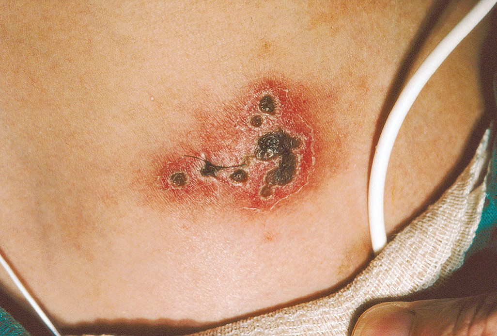
Erythematous plaque with necrosis and eschar formation in a young female with immunosuppression and disseminated Aspergillus fumigatus infection.

(From Paller AS, Mancini AJ: Hurwitz clinical pediatric dermatology, a textbook of skin disorders of childhood and adolescence, ed 5, Philadelphia, 2016, Elsevier.)

Figure E5 Aspergillosis vertebral osteomyelitis and diskitis.
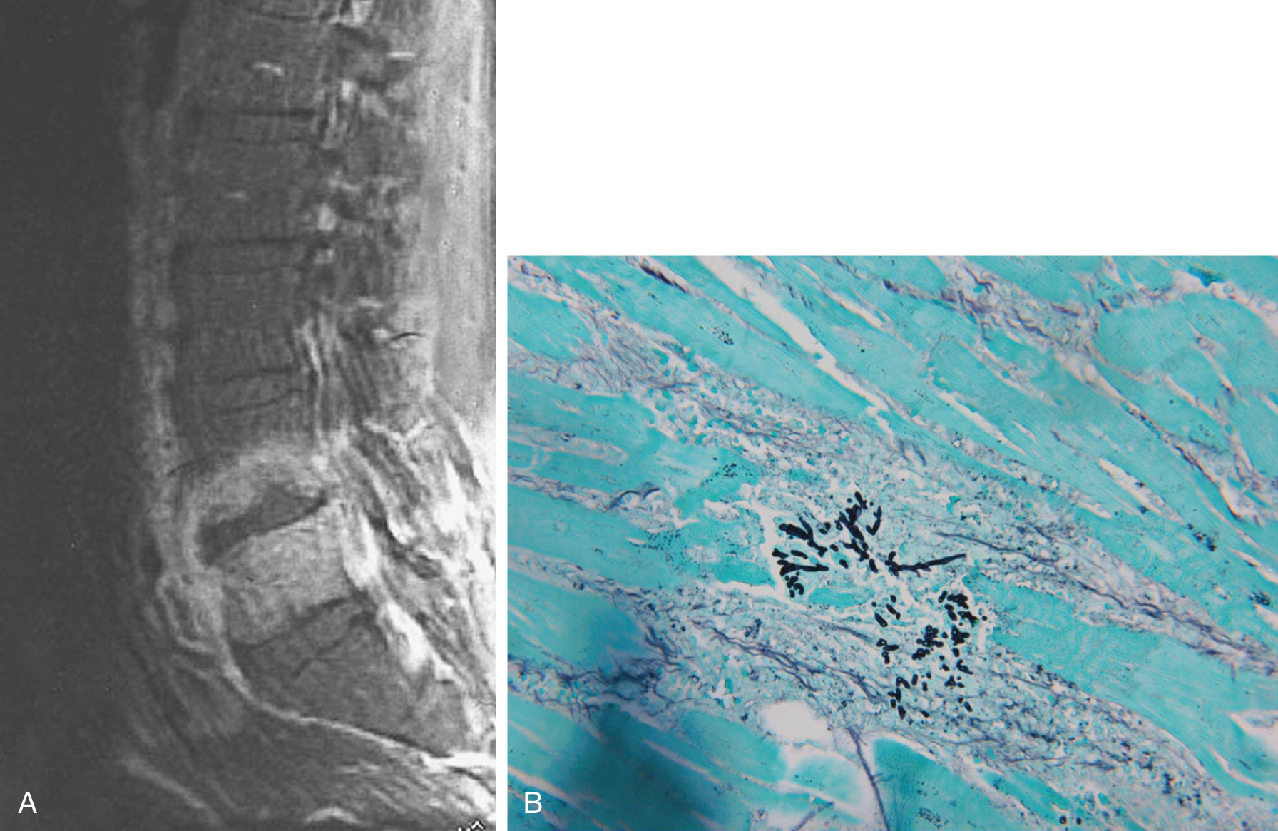
(A)Aspergillus may spread directly from the lung to adjacent vertebrae, disk spaces, and ribs (more often in children) or through the bloodstream. (B) Infected tissue may show characteristic organisms.

(From Firestein GS et al: Firestein & Kelley’s textbook of rheumatology, ed 12, Philadelphia, 2024, Elsevier.)

Diagnosis ⬆ ⬇

Differential Diagnosis

  • •Tuberculosis
  • •Cystic fibrosis
  • •Carcinoma of the lung
  • •Eosinophilic pneumonia
  • •Bronchiectasis
  • •Sarcoidosis
  • •Lung abscess
Workup

Physical examination and laboratory data

Laboratory Tests
Abpa:

  • •Peripheral blood eosinophilia and an elevated total serum immunoglobulin E (IgE) level
  • •Immediate skin test reactivity with Aspergillus spp2
  • •Aspergillus serum precipitating antibody
  • •Sputum cultures may be positive for Aspergillus spp, but are nonspecific and may represent colonization or contamination depending on the clinical context
  • •Diagnostic criteria for ABPA are summarized in Table E3

TABLE E3 Diagnostic Criteria for Allergic Bronchopulmonary Aspergillosis

Newly Proposed Diagnostic Criteria
Predisposing conditions
  • •Asthma
  • •Central bronchiectasis (inner two thirds of chest CT field)
  • •COPD
Essential criteria (both should be present)*
  • •Total serum IgE concentration >500 IU/ml

  1. A. fumigatus-specific IgE ≥0.35 kUA/L or positive skin testing
Other criteria (at least two of three)
  1. 1.Positive IgG against A fumigatus
  2. 2.Blood eosinophil count ≥500 cells/μL
  3. 3.CT consistent with ABPA (bronchiectasis, mucus plugging, high-attenuation mucus or fleeting opacities consistent with ABPA)

ABPA, Allergic bronchopulmonary aspergillosis; CT, computed tomography; IgE, immunoglobulin E; IU, international units.

* CT with high-attenuation mucus is diagnostic of ABPA even if other criteria are not met.

From Agarwal R et al: Revised clinical practice guidelines for diagnosing, classifying, and treating allergic bronchopulmonary aspergillosis/mycoses: a Delphi statement from the ISHAM-ABPA working group, Eur Resp J 63(4):2400061, 2024. doi: https://doi.org/10.1183/13993003.00061-2024. Table 1.

Aspergillomas:

  • •Sputum culture
  • •Serum precipitating antibody
Invasive Aspergillosis:

  • •Definitive diagnosis requires the demonstration of tissue invasion (i.e., septate, acute angle branching hyphae) or a positive culture from the tissue or fluid obtained by an invasive procedure such as transbronchial biopsy.
  • •Sputum and nasal cultures: In high-risk patients a positive culture is strongly suggestive of invasive aspergillosis.
  • •Serology: The Platelia Aspergillus ELISA assay detects a circulating fungal antigen, galactomannan. The galactomannan antigen immunoassay is often used for diagnosis. Galactomannan is a polysaccharide contained in the cell wall of Aspergillus. Its presence in serum or other body fluids, such as samples obtained from bronchoalveolar lavage, is indicative of invasive infection, and it is recommended as an accurate marker for diagnosis in certain patient subpopulations (hematologic malignancy and hematopoietic stem-cell transplantation). The sensitivity of the galactomannan antigen immunoassay can be impaired by antifungal therapy. The β-D glucan assay can also be used to detect early infection but is not specific for Aspergillus species.5
  • •The sensitivity of the galactomannan assay for invasive pulmonary aspergillosis is higher in bronchoalveolar lavage fluid than in serum. In patients with risk factors and radiologic findings suggestive of invasive aspergillosis, a positive galactomannan confirms the diagnosis of probable invasive pulmonary aspergillosis.10
  • •Blood cultures: Usually negative (Aspergillus is rarely found in the blood).
  • •Lung biopsy is necessary for definitive diagnosis.
  • •Biopsy and culture of extrapulmonary lesions.
  • •Polymerase chain reaction assays and next generation sequencing from bronchoalveolar lavage samples may be employed in a case-by-case basis, but their results should be interpreted in conjunction with other diagnostic tests and the clinical context.
Imaging Studies
Abpa:

  • •CXRs show a variety of abnormalities, from small, patchy, fleeting infiltrates (commonly in the upper lobes) to lobar consolidation or cavitation.
  • •A majority of patients eventually develop central bronchiectasis.1
Aspergillomas:

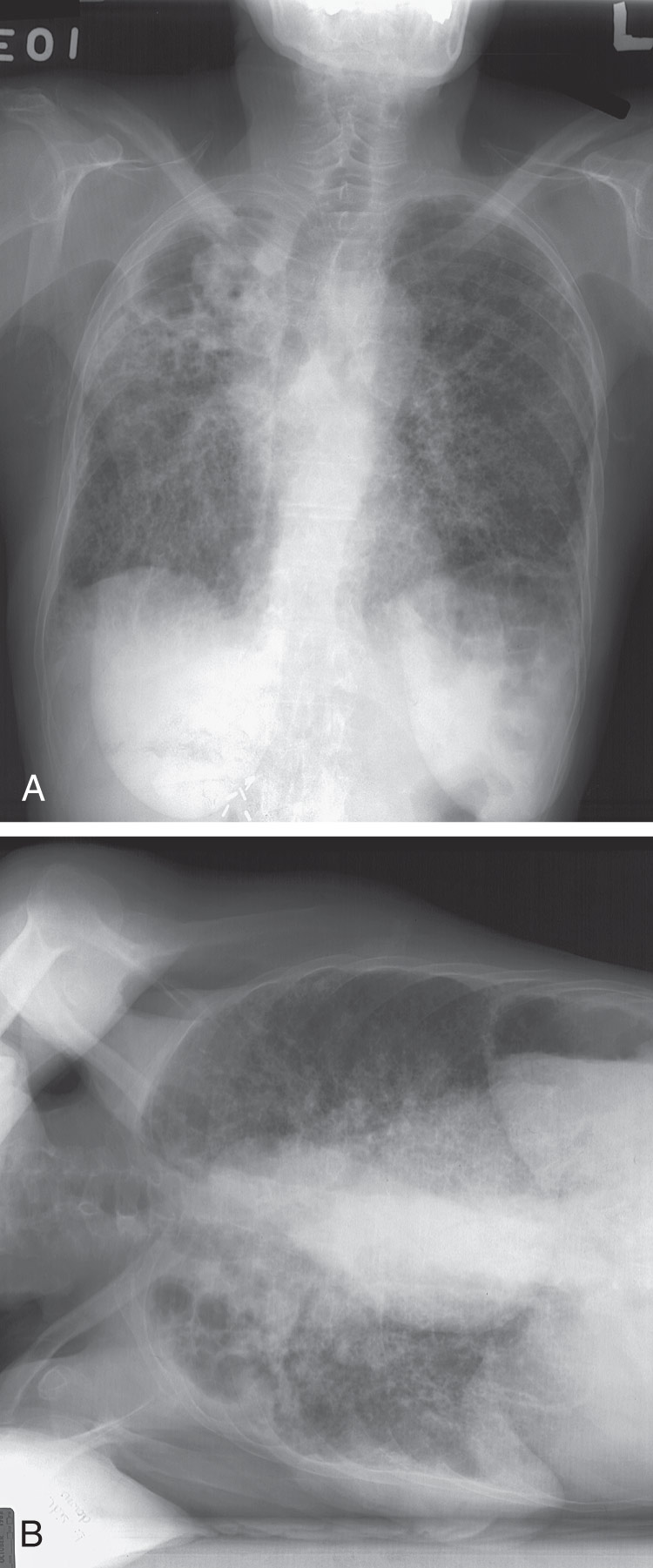
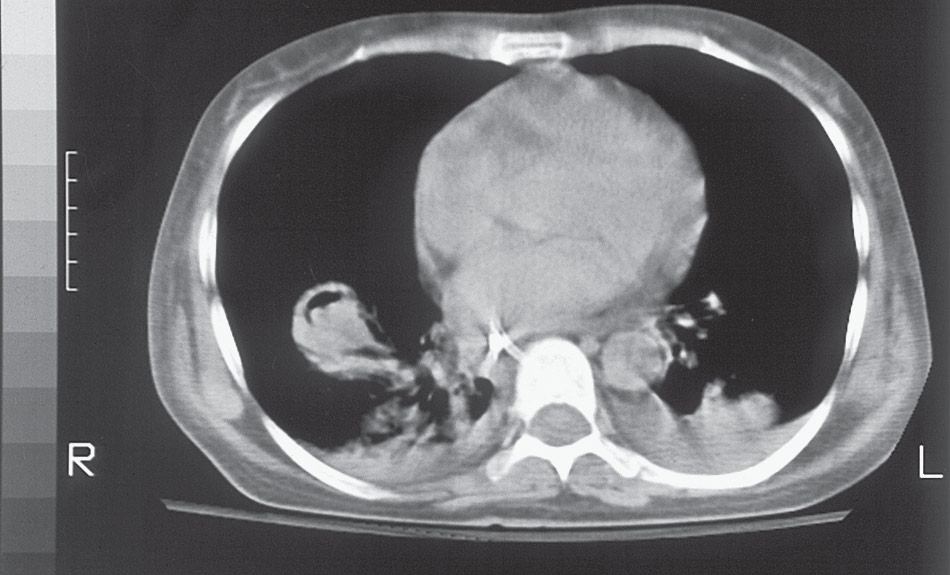
  • •CXR or CT scans usually show the characteristic intracavity mass (Figs. E6 andE7) partially surrounded by a crescent of air.

Figure E6 Computed Tomography Scan Demonstrating an Aspergilloma Involving the Right Lower Lobe

(From Sellke FW et al: Sabiston and Spencer: surgery of the chest, ed 10, Philadelphia, 2024, Elsevier.)

Figure E7 Posteroanterior (A) and Decubitus (B) Chest Radiographs Demonstrating an Aspergilloma Within a Right Upper Lobe Cavity

The fungus ball "moves" with change in patient position.

(From Sellke FW et al: Sabiston and Spencer: surgery of the chest, ed 10, Philadelphia, 2024, Elsevier.)

Invasive Aspergillosis:

  • •Chest CT scan may reveal cavity formation and the "halo sign" (ground glass opacity surrounding a nodule that is very suggestive of angioinvasive disease). May also see dense nodules, which may cavitate.

Treatment ⬆ ⬇

Acute General Rx
Abpa:

  • •Prednisone (0.5 to 2 mg/kg/day PO) for 1 to 2 wk, followed by a taper over 2 to 3 mo plus oral itraconazole 5 mg/kg/day up to 400 mg/day with therapeutic drug monitoring for a course of 3 to 6 mo. Alternatives: Voriconazole or posaconazole may be effective.2
  • •If a patient is corticosteroid dependent, prophylaxis for the prevention of Pneumocystis jiroveci infection and maintenance of bone mineralization should be considered.
  • •Bronchodilators and physiotherapy.
  • •Serial CXR and serum IgE may be useful in guiding treatment.
Aspergillomas:

  • •Controversial and problematic; the optimal treatment strategy is unknown.
  • •Up to 10% of aspergillomas may resolve clinically without overt pharmacologic or surgical intervention.
  • •Observation for asymptomatic patients.
  • •Surgical resection/arterial embolization for those patients with severe hemoptysis or life-threatening hemorrhage.
  • •For those patients at risk for marked hemoptysis with inadequate pulmonary reserve, consider voriconazole 6 mg/kg IV/PO bid on day 1; then 4 mg/kg IV/PO bid. Isavuconazonium sulfate or itraconazole may also be used as well as transthoracic, intracavitary installation of amphotericin B in poor surgical candidates. (Note: Aspergillus now has a higher rate of resistance to itraconazole.)
Invasive Aspergillosis:

  • •Voriconazole 6 mg/kg IV/PO bid on day 1 followed by 4 mg/kg IV/PO bid. Voriconazole serum concentrations need to be monitored to achieve a target range of 1.0 to 5.5 mg/L (trough on day 4).
  • •Isavuconazonium sulfate (prodrug of isavuconazole) 372 mg IV/PO three times a day for six doses and then 372 mg IV/PO daily.
  • •Posaconazole: May be administered as delayed-release tabs (300 mg PO two times on day 1, then 300 mg PO daily) or suspension (less preferred; 200 mg PO four times on day 1, then 400 mg PO twice a day after stabilization of disease) or intravenously (300 mg IV over 90 min twice on day 1, then 300 mg IV daily). Posaconazole serum concentrations also may be monitored.
  • •Posaconazole may also be used as prophylaxis against invasive disease in certain high-risk populations, such as neutropenic patients with acute myeloid leukemia or those with graft versus host disease.
  • •Antifungal agents for invasive aspergillosis are summarized in Table E4.

TABLE E4 Antifungal Agents for Invasive Aspergillosis

AgentClassRoute of AdministrationDoseComments
Primary Therapy
VoriconazoleAzoleIV/PO6 mg/kg (IV) q12h × 2 doses, followed by 4 mg/kg (IV) q12h or 200 mg (PO) q12h. Some experts advise oral dosing as 4 mg/kg (PO) q12h.Recommended for primary therapy in most patients; 4,12 caution for use in patients with potential liver toxicity and for drug interactions; measurement of serum levels for efficacy and avoidance of toxicity13
Alternative Primary Therapy
IsavuconazoleAzoleIV/PO372 mg q8h × 6 doses, IV or PO followed by 372 mg daily (372 mg isavuconazonium sulfate = 200 mg isavuconazole)Well tolerated, efficacy in treatment of pulmonary aspergillosis, and fewer adverse events than voriconazole-treated patients14
PosaconazoleAzoleIV/POTablet or IV, 300 mg q12h × 2 doses, followed by 300 mg dailyOral and IV formulation; noninferior to voriconazole for treatment;15-used for prophylaxis and treatment
Liposomal amphotericin BPolyeneIV3-5 mg/kg/dayWell tolerated; minimal infusion reactions or nephrotoxicity compared to prior formulations; initial doses of 10 mg/kg/day more toxic and not more effective. Reserved for patients with resistance or those unable to tolerate azoles16
Other Agents
ItraconazoleAzolePO200 mg twice daily (PO) or 65 mg twice daily of SUBA-itraconazoleErratic bioavailability improved with oral solution and capsule, serious cardiac adverse events with higher doses, drug interactions common; IV formulation not currently available17
CaspofunginEchinocandinIV70 mg first day then 50 mg/dayApproved for refractory infection and intolerance to standard therapy; well tolerated;18 limited efficacy as monotherapy for primary infection19
MicafunginEchinocandinIVInvestigational for Aspergillus treatment (U.S.) (50-100 mg/day)Efficacy for prevention and salvage treatment of aspergillosis20,21
AnidulafunginEchinocandinIVInvestigational for Aspergillus (200 mg first day then 100 mg/day)Combination trial data showing improved outcomes in patients with galactomannan diagnosis of invasive pulmonary aspergillosis22

From Bennett JE et al: Mandell, Douglas, and Bennett’s principles and practice of infectious diseases, ed 9, Philadelphia, 2020, Elsevier.

Alternative Treatment:

  • •Amphotericin B lipid complex (ABLC) 5 mg/kg IV daily
  • •Liposomal amphotericin B (L-AMB) 3 to 5 mg/kg IV daily
  • •Because azoles and echinocandins target different cellular sites, combination therapy may have additive activity against Aspergillus species
  • •Investigational antifungals in clinical trials with novel mechanisms of action include olorofim, fosmanogepix, and aerosolized opelconazole. Encochleated amphotericin B oral formulation is also under investigation
Referral

To an infectious diseases specialist

Pearls & Considerations ⬆ ⬇

BOX E1 Use of Antifungal Agents in Combination

The development of new antifungal drugs gives the clinician more options for prophylaxis and therapy than in previous years. There is an overall level of simplicity to the drug choices once their mechanisms of action are understood. The polyenes, including amphotericin products and the topical agent nystatin, attach onto ergosterol in the fungal cell membrane and are considered fungicidal because cytoplasm leaks out and individual cells die. The azoles, including fluconazole, itraconazole, voriconazole, and posaconazole, prevent the formation of new ergosterol. Azoles are considered fungistatic because removal of the drug permits cell regrowth. Theoretically, use of an azole together with a polyene may have an overall static effect for an established infection as the ergosterol target for the fungicidal polyene is depleted. However, this combination may have advantages in terms of enhanced spectrum of activity. The echinocandins, including caspofungin, micafungin, and anidulafungin, prevent interaction of the catalytic and regulatory subunits of the β-glucan synthesis enzyme, so less β-glucan is formed for the cell wall. The scaffolding for the fungal cell wall is not maintained, and a dividing cell may burst open when trying to extend the new cell wall over daughter cells. The echinocandins are considered fungicidal for yeasts but fungistatic for molds because drug activity is concentrated at only the tips of the extending hyphae with little effect on less metabolically active subapical compartments of the fungus. Combination therapy may have the most effect when a cell wall agent (an echinocandin) is used together with a cell membrane agent (a polyene or an azole). There is no role for three-drug therapy (an echinocandin, a polyene, and an azole). Aside from cases of cryptococcal meningitis, in which the importance of combination therapy is well established, the benefits of frontline use of combination antifungal for molds remains controversial, although active investigation continues in clinical trials. The value of combination regimens as salvage therapy for refractory mold infections remains uncertain.

From Hoffman R et al: Hematology, basic principles and practice, ed 8, Philadelphia, 2023, Elsevier.

Related Content

Aspergillosis (Patient Information)

Reference(s) ⬆

  1. Greenberger PA : Allergic bronchopulmonary aspergillosishttp://dx.doi.org/10.1067/mai.2002.130179J Allergy Clin Immunol. 110(5):685-692, 2002.
  2. Thompson GR, Patterson TF : Chapter 257: Aspergillus species Bennett JE, Dolin R, Blaser MJ, editors : Mandell, Douglas, and Bennett’s principles and practice of infectious diseases. ed 9Elsevier-Philadelphia, PA:3103-3116, 2020.
  3. Cunha C : PTX3 deficiency and aspergillosisN Engl J Med. 370(17):1666-1667, 2014.
  4. Ghex D : Early-onset invasive aspergillosis and other fungal infections in patients treated with ibrutinibdoi:10.1182/blood-2017-11-81286Blood. 131(17):1955-1959, 2018.
  5. Patterson TF : Practice guidelines for the diagnosis and management of aspergillosis: 2016 update by the Infectious Diseases Society of AmericaClin Infect Dis. 63(4):e1-e60, 2016.
  6. Chong WH, Neu KP : Incidence, diagnosis and outcomes of COVID-19- associated pulmonary aspergillosis (CAPA): a systematic reviewJournal of Hospital Infection. 113:115-129, 2021.
  7. Garcia-Vidal C : Invasive aspergillosis complicating pandemic influenza A (H1N1) infection in severely immunocompromised patientshttp://dx.doi.org/10.1093/cid/cir485Clin Infect Dis. 53(6):e16-e19, 2011.
  8. Crum-Cianflone NF : Invasive aspergillosis associated with severe influenza infectionsOpen forum Infect Dis. 3(3):ofw171, 2016.
  9. Walsh TJ : Chapter 319: Aspergillosis Goldman L, Schafer A, editors : Goldman-Cecil medicine. ed 26Elsevier-Philadelphia, PA:2052-2056, 2020.
  10. Cook RJ : Aspergillus infection of implantable cardioverter-defibrillatorhttp://dx.doi.org/10.4065/79.4.549Mayo Clin Proc. 79(4):549-552, 2004.
  11. Buil JB : Trends in azole resistance in Aspergillus fumigatus, the Netherlands 1994-2016Emerg Infec Dis. 25(1):176-178, 2019.
  12. Denning DW : Efficacy and safety of voriconazole in the treatment of acute invasive aspergillosisClin Infect Dis. 34:563-571, 2002.
  13. Pascual A : Challenging recommended oral and intravenous voriconazole doses for improved efficacy and safety: population pharmacokinetics-based analysis of adult patients with invasive fungal infectionsClin Infect Dis. 55:381-390, 2012.
  14. Maertens JA : Isavuconazole versus voriconazole for primary treatment of invasive mould disease caused by Aspergillus and other filamentous fungi (SECURE): a phase 3, randomised-controlled, non-inferiority trialLancet. 387:760-769, 2016.
  15. Maertens JA : Posaconazole versus voriconazole for primary treatment of invasive aspergillosis: a phase 3, randomised, controlled, non-inferiority trialThe Lancet. 397.10273:499-509, 2021.
  16. Cornely OA : Liposomal amphotericin B as initial therapy for invasive mold infection: a randomized trial comparing a high-loading dose regimen with standard dosing (AmBiLoad trial)Clin Infect Dis. 44:1289-1297, 2007.
  17. Patterson TF : Invasive aspergillosis. Disease spectrum, treatment practices, and outcomes. I3 Aspergillus Study GroupMedicine (Baltimore). 79:250-260, 2000.
  18. Maertens J : Efficacy and safety of caspofungin for treatment of invasive aspergillosis in patients refractory to or intolerant of conventional antifungal therapyClin Infect Dis. 39:1563-1571, 2004.
  19. Herbrecht R : Caspofungin first-line therapy for invasive aspergillosis in allogeneic hematopoietic stem cell transplant patients: an European Organisation for Research and Treatment of Cancer studyBone Marrow Transplant. 45:1227-1233, 2010.
  20. Marr KA : Combination antifungal therapy for invasive aspergillosisClin Infect Dis. 39:797-802, 2004.
  21. Denning DW : Micafungin (FK463), alone or in combination with other systemic antifungal agents, for the treatment of acute invasive aspergillosisJ Infect. 53:337-349, 2006.
  22. Marr KA : Combination antifungal therapy for invasive aspergillosis: a randomized trialdoi:https://doi.org/10.7326/M13-2508Ann Int Med. 162(2):81-89, 2015.