section name header

Basic Information ⬇

Author: Glenn G. Fort, MD, MPH

Definition

Infection with cytomegalovirus (CMV), a herpes virus, is common in the general population, with multiple mechanisms for transmission, often during childhood and adolescence. CMV is associated with pregnancy and can be a congenital disease. CMV is also associated with immunocompromised states and may be life-threatening.

Synonyms

  • CMV
  • Heterophil-negative mononucleosis
  • Cytomegalic inclusion disease virus
ICD-10CM CODES
B25.9Cytomegaloviral disease, unspecified
P35.1Congenital cytomegalovirus infection
Z20.820Contact with and (suspected) exposure to varicella
Epidemiology & Demographics

  • •Seroprevalence is widespread: 60% to 90% antibody positivity in adults.
  • •Increased infection develops perinatally, in day care exposure, and then during reproductive age, related to sexual activity.
Routes Of Transmission

  • •Blood transfusions
  • •Sexually transmitted diseases (STDs) via uterus, cervix, and semen
  • •Perinatally via breast milk
  • •Transplant of organs-bone marrow, kidneys, liver, heart, or lung
  • •Saliva
Physical Findings & Clinical Presentation
Children:

Congenital-25% of infected children with symptoms if congenital (Table E1):

  • •Petechial rash
  • •Jaundice and/or hepatosplenomegaly
  • •Lethargy
  • •Respiratory distress
  • •Central nervous system (CNS) involvement, seizures

TABLE E1 Findings in Infants With Symptomatic Congenital Cytomegalovirus Infection

Findings% of Infants
Clinical Findings
Prematurity (<37 wk)24
Jaundice (direct bilirubin >2 mg/dl)42
Petechiae54
Hepatosplenomegaly19
Purpura3
Microcephaly35
IUGR28
One clinical finding41
Two clinical findings59
Laboratory Findings
Elevated ALT (>80 IU/ml)71
Thrombocytopenia (<100,000 k/mm2)43
Direct hyperbilirubinemia (>2 mg/dl)54
Head CT abnormalities42

Findings in 70 infants with symptomatic congenital CMV infection identified during newborn screening program for infants with congenital CMV infection at the University of Alabama Hospitals over an approximate 20-yr interval.

ALT, Alanine aminotransferase; CMV, cytomegalovirus; IUGR, in utero growth restriction.

From Kliegman RM et al: Nelson textbook of pediatrics, ed 22, Philadelphia, 2025, Elsevier.

Postnatal acquisition:

  • •CMV mononucleosis
  • •Pharyngitis, croup, bronchitis, pneumonia
Healthy Adults:

Common:

  • •May be asymptomatic
  • •CMV mononucleosis similar to Epstein-Barr virus (EBV) mononucleosis
  • •Fever-lasting 9 to 30 days-mean of 19 days

Less common:

  • •Exudative pharyngitis
  • •Lymphadenopathy, hepatitis, splenomegaly
  • •Interstitial pneumonia (rare)
  • •Nonspecific rash
  • •Thrombocytopenia/hemolytic anemia

Rare:

  • •Guillain-Barré syndrome
  • •Meningoencephalitis
  • •Myocarditis
Immunosuppressed Patients:

  • •Febrile mononucleosis
  • •GI ulcerations, hepatitis, pneumonitis, retinitis, encephalopathy, meningoencephalopathy
  • •HIV associated-dementia, demyelination, retinitis (Fig. E1), acalculous cholecystitis, adrenalitis, diarrhea, enterocolitis, esophagitis
  • •Diabetes associated with pancreatitis
  • •Adrenalitis associated with HIV
  • •Findings in CMV infections in solid organ transplant recipients are described in Table E2

Figure E1 CMV retinitis (hemorrhages and inflammation).
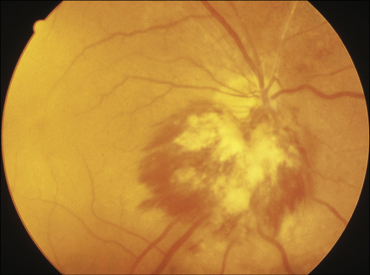
(From Zamir E: Ocular infections with cytomegalovirus. In Yanoff M, Duker JS [eds]: Ophthalmology, ed 4, 2014, Elsevier, in Spec, A et al: Comprehensive review of infectious diseases, 2020, Elsevier.)

TABLE E2 Findings in Cytomegalovirus Infections in Solid Organ Transplant Recipients

Clinical FindingsLaboratory Findings
CMV Syndrome
Fever, nonspecific findings, fatigueLeukopenia, thrombocytopenia, reactive lymphocytosis, hepatitis, CMV DNA in blood
End-Organ Disease
Gastrointestinal disease, including esophagitis, colitis, and hepatitisDetection of CMV DNA in blood; detection of CMV in tissue biopsy; hepatitis, including elevated bilirubin
Lung disease; hypoxemiaAbnormalities in lung imaging
CMV in bronchoalveolar lavage fluid
EncephalitisCSF pleocytosis, elevated CSF protein
Abnormalities in CNS imaging
Allograft dysfunctionEvidence of graft rejection

From Kliegman RM et al: Nelson textbook of pediatrics, ed 22, Philadelphia, 2025, Elsevier.

Etiology

  • •CMV is the fifth member of the human herpes family: Human herpesvirus (HHV)-5. It is a double-stranded DNA virus and has an icosahedral shape measuring 150 to 200 nm in diameter.1
  • •CMV infection can remain latent and reactivate with immunosuppression.

Diagnosis ⬆ ⬇

Differential Diagnosis

Congenital:

  • •Acute viral, bacterial, parasitic infections including other congenitally transmitted agents (toxoplasmosis, rubella, syphilis, pertussis, croup, bronchitis)

Acquired:

  • •EBV mononucleosis
  • •Viral hepatitis-A, B, C
  • •Cryptosporidiosis
  • •Toxoplasmosis
  • •Mycobacterium avium infections
  • •Human herpesvirus 6
  • •Acute HIV infection
Workup

  • •Laboratory confirmation (Table E3) combined with clinical findings often with leukopenia, thrombocytopenia, lymphocytosis. Diagnostic modalities include serologic assays, PCR, detection of CMV PP25 antigen in leukocytes, isolation of virus from body fluids and urine, and cytopathic demonstration of "owl eye" intracellular inclusions
  • •Serology:
    1. 1.Detection of CMV-IgM antibodies suggests recent infection. CMV-IgG antibodies usually appear 2 to 3 wk after infection.
    2. 2.Molecular assays (polymerase chain reaction [PCR] viral loads): On plasma.
    3. 3.CMV antigenemia assays: Detects antibodies to the pp65 protein of the virus in peripheral blood leukocytes. These tests and the PCR tests are used in immunocompromised, AIDS, and transplant patients.
  • •Cultures: Using human fibroblast cultures of blood, cerebrospinal fluid, urine, bronchoalveolar lavage, and biopsy specimens but can take 1 to 6 wk
  • •Fundoscopic: Necrotic patches with white granular component of retina
  • •Biopsy: "Owl eye" inclusion bodies on tissue sample
  • •HIV

TABLE E3 Laboratory Detection of Cytomegalovirus Infection

AssayApplicationCharacteristics of Assay
Tissue Culture
Virus isolationDetection of virus in clinical materialRequires prolonged culture periods; low sensitivity compared with molecular techniques; expensive
Histopathology
HistopathologyIdentification of CMV-infected cells (owl’s eye inclusions)Routine for most laboratories; low sensitivity is improved with IHC (see below)
Viral antigen detection (immunohistochemistry)Sensitive and allows detection of CMV-infected cells in variety of specimensRapid and can be quantitative
Molecular Methodologies
Nucleic acid amplification testing (NAAT), including quantitative PCR, transcription-mediated amplification (TMA)Quantitation of CMV DNA and viral RNARapid, sensitive, and quantitative; can be applied to variety of clinical specimens (blood, saliva, urine, biopsy specimens, etc.)
DNA/RNA in-situ hybridizationSensitive detection of CMV DNA or RNA transcripts in tissue specimensTechnically more difficult than NAAT, long turnaround times, can identify specific cells infected with CMV
Serologic Testing
CMV-specific IgM serologyScreening for recent infectionsVariability in duration of response and low levels of IgM antibodies decrease sensitivity and specificity
CMV-specific IgG serologyDetection of infection with HCMV, useful for seroprevalence studiesRapid, specific, and quantitative; gold standard for CMV infection (acute and past)
CMV-specific IgG avidityEstimate of duration of HCMV infectionInterpretation limited to high and low values of avidity; useful for timing of recent CMV infections in pregnancy; not useful in allograft recipients

CMV, Cytomegalovirus; DNA, deoxyribonucleic acid; HCMV, human cytomegalovirus; IgG, immunoglobulin G; PCR, polymerase chain reaction; RNA, ribonucleic acid.

From Kliegman RM et al: Nelson textbook of pediatrics, ed 22, Philadelphia, 2025, Elsevier.

Imaging Studies

  • •Chest x-ray examination: If pneumonitis suspected, consider bronchoscopy
  • •Endoscopy: If GI involvement
  • •Computed tomography scan/MRI: If CNS involvement

Treatment ⬆ ⬇

Nonpharmacologic Therapy

  • •Strict handwashing and standard precautions limit CMV transmission in health care facilities.
  • •Antiretroviral therapy (ART) in patients with CD4 count <50/mm2 for the goal of CD4 >100/mm2 for a 3- to 6-mo period.
Acute General Rx

Antiviral therapy with intravenous ganciclovir or oral valganciclovir is appropriate in immunocompromised patients. It can also be used in immunocompetent patients with severe disease.

For compromised hosts with CMV retinitis or pneumonitis:

  • •Ganciclovir 5 mg/kg q12h intravenous (IV) × 14 to 21 days, then valganciclovir: 900 mg PO q24h or alternative regimen
  • •Ganciclovir intraocular implant plus valganciclovir 900 mg PO q24h or alternative regimen
  • •Foscarnet 90 mg/kg q12h × 14 to 21 days, then 90 mg to 120 mg/kg IV q24h or alternative regimen
  • •Cidofovir 5 mg/kg IV q day × 14 days, then 5 mg/kg IV q2wk
  • •Fomivirsen-salvage therapy for CMV retinitis 300 μg injected into vitreous
  • •Letermovir, in IV and oral form, is approved for prophylaxis only of CMV infection and disease in seropositive recipients of an allogenic hematopoietic stem cell transplant: 480 mg IV or PO q24h. However, this is very expensive. Suggestions for management of possible CMV infection after HSCT are summarized in Table E4.
  • •Maribavir is indicated for the treatment of adults and pediatric patients ( ≥12 yr of age, weight ≥35 kg) with posttransplant cytomegalovirus infection/disease that is refractory to treatment to other agents. Dose: 400 mg po twice daily, with or without food.3

TABLE E4 Suggestions for Management of Possible CMV Infection After HSCT

IndicationStrategyaComment
Prevention
Allogeneic Transplant
Seropositive recipientPreemptive ganciclovirb induction for subclinical viremia, 5 mg/kg bid for 7-14 days, followed by 5 mg/kg daily until the end of maintenance or ganciclovir prophylaxisc at engraftmentSome cases of CMV disease may occur shortly after ganciclovir discontinuation.
CMV reactivation might be delayed, occurring later after HSCT.
Seronegative recipient with seropositive donorPreemptive ganciclovir induction for subclinical viremia, 5 mg/kg bid for 7-14 days, followed by 5 mg/kg daily until the end of maintenance and seronegative or filtered blood productsProphylaxis at engraftment is not recommended because of the low incidence of posttransplantation infection.
Seronegative recipient with seronegative donorSeronegative or filtered blood products
Autologous Transplant
Seropositive recipientEarly ganciclovir induction of subclinical viremia, 5 mg/kg ganciclovir bid for 7 days, followed by 5 mg/kg daily for 14 days of maintenanceBecause of the very low risk in some settings, monitoring is not uniformly advocated.
Seronegative recipientSeronegative or filtered blood products
Treatment of Disease
CMV pneumonitisGanciclovir induction, 5 mg/kg bid for 14-21 days, followed by 5 mg/kg daily for at least 3-4 wk of maintenance plus IVIG every other day for the duration of inductionExtended maintenance throughout periods of severe immunosuppression (i.e., GVHD treatment) may be considered.
Gastrointestinal diseaseGanciclovir induction, 5 mg/kg bid for 14-21 days, followed by 5 mg/kg daily for at least 3-4 wk of maintenanceIf deep ulcerations are present, maintenance may be required for a longer time.
Marrow failureFoscarnet, 90 mg/kg bid for 14 days, followed by 90 mg/kg daily for 2 wk plus G-CSFGanciclovir plus IVIG has also been used.
RetinitisGanciclovir, 5 mg/kg bid for 14-21 days, followed by 5 mg/kg daily for at least 3-4 wkExtended maintenance may be required.

bid, Twice daily; CMV, cytomegalovirus; G-CSF, granulocyte colony-stimulating factor; GVHD, graft-versus-host disease; HSCT, hematopoietic stem cell transplantation; IVIG, intravenous immune globulin; PCR, polymerase chain reaction.

a Regimens should be accompanied by weekly monitoring with antigenemia or PCR-based nucleic acid testing.

b Oral 900-mg doses of valganciclovir produce blood levels that are similar to those for the standard intravenous dose (5 mg/kg) of ganciclovir. Foscarnet and cidofovir are acceptable alternatives. Renal dose adjustment is required for all antiviral agents.

c Foscarnet, high-dose acyclovir, or valacyclovir are acceptable alternatives. Renal dose adjustment required for all antiviral agents.

From Bennett JE et al: Mandell, Douglas, and Bennett’s principles and practice of infectious diseases, ed 9, Philadelphia, 2020, Elsevier.

Disposition

  • •CMV infection in patients who are immunocompromised (especially those with AIDS, bone marrow and solid organ transplant recipients, and disorders of cell-mediated immune function) will need expert, long-term follow-up by an infectious disease specialist or immunologist familiar with the care of such patients.
  • •CMV mononucleosis, hepatitis, pharyngitis, etc. in immunologically normal hosts are usually self-limiting infections requiring no special follow-up plans.
Referral

  • •To an ophthalmologist if CMV retinitis is present
  • •To an infectious disease specialist or AIDS specialist for patients who are HIV-positive with CMV disease
  • •To a cellular immunologist or transplant specialist in the case of CMV infection in a transplant recipient
  • •To a pediatric infectious disease specialist for congenital CMV infection

Pearls & Considerations ⬆ ⬇

Reference(s) ⬆

  1. Dioverti MV, Razonable RR : Cytomegalovirushttps://doi.org/10.1128/microbiolspec.dmih2-0022-2015Microbiol Spectr. 4(4), 2016.
  2. Hughes BL : A trial of hyperimmune globulin to prevent congenital cytomegalovirus infectionNEJM. 385:436-444, 2021.
  3. Imlay HN, Kaul DR : Letermovir and maribavir for the treatment and prevention of cytomegalovirus infection in solid organ and stem cell transplant recipientsClin Infect Dis. 73:156-160, 2021.