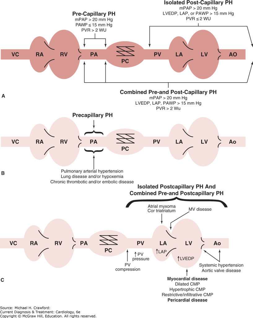
The clinical classification of PH was updated in 2022 (Table 30–1). A hemodynamic approach to classification of PH is illustrated in Figure 30–1. PH severity is graded by the value of the mPAP: mild (20–40 mm Hg), moderate (41–55 mm Hg), or severe (> 55 mm Hg).
Table 30–1. Classification System for Pulmonary Hypertension
Group 1: Pulmonary arterial hypertension
Group 2: Pulmonary hypertension due to left heart disease
Group 3: Pulmonary hypertension due to lung disease and/or hypoxia
Group 4: pH due to pulmonary artery obstructions
Group 5: Pulmonary hypertension with unclear multifactorial mechanisms
|
Figure 30-1. Hemodynamic classification of pulmonary hypertension (PH). A: Pulmonary hypertension may result from pathology in precapillary or postcapillary pulmonary circulation. B: Common causes of precapillary PH. C: Common causes of postcapillary and mixed PH. Ao, aorta; CMP, cardiomyopathy; DTPG, diastolic transpulmonary gradient; LA, left atrium; LV, left ventricle; LVEDP, left ventricular end-diastolic pressure; PA, pulmonary artery; PAWP, pulmonary arterial wedge pressure; PC, pulmonary capillaries; PV, pulmonary veins; PVR, pulmonary vascular resistance; RA, right atrium; RV, right ventricle; TPG, transpulmonary pressure gradient; VC, vena cava. (Reproduced with permission from De Marco T, et al. Pulmonary hypertension in left heart disease—systolic, diastolic, valvular. In: Kirklin JK (ed), Pulmonary Hypertension and Right Heart Failure: ISHLT Monograph Series. Vol 9. 2015.)
A. Pulmonary Arterial Hypertension
Pathologic abnormalities in PAH are localized to the precapillary pulmonary arterioles, resulting in a characteristic hemodynamic profile that can be identified during right heart catheterization (RHC) (see Figure 30–1). Hemodynamically, PAH is defined by a sustained elevation of the mPAP ≥ 20 mm Hg and a PAWP or left ventricular end-diastolic pressure ≤ 15 mm Hg (normal PAWP, 8–12 mm Hg) and a PVR >2 Wood units (WU).
PAH is caused by a disparate group of diseases that have in common vasoconstriction and arteriopathy with remodeling of the precapillary pulmonary arterioles (see Table 30–1). Chronically elevated PAP leads to right ventricular failure and death. Although the prognosis for patients with PAH varies with the underlying cause, overall survival is poor even with modern therapies.
The prevalence of PAH in the United States is estimated to be between 50,000 and 100,000 cases. Of these, only 15,000 to 25,000 cases are appropriately diagnosed and treated. Multicenter registry data suggest that most patients with PAH are women (with a 2:1 female-to-male ratio), with a mean age of 50 years. Seventy-five percent of patients with PAH have symptoms at rest or with minimal exertion. The mean duration of symptoms prior to diagnosis is 24 months.
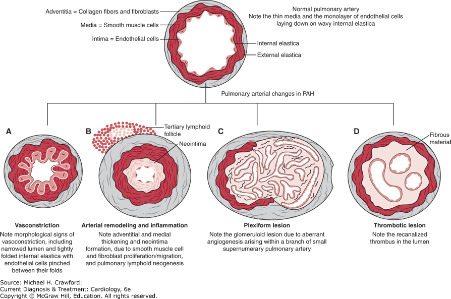
Three processes contribute to pulmonary artery luminal narrowing in PAH: vasoconstriction, arterial remodeling, and thrombosis in situ (Figure 30–2). In susceptible individuals, pulmonary vascular injury leads to disruption of the homeostatic balance present in the healthy pulmonary arteries. Upregulation of vasoconstrictive and proproliferative mediators (ie, endothelin-1, serotonin, and thromboxane) and downregulation of vasodilatory and antiproliferative mediators (ie, nitric oxide, prostacyclin, and smooth muscle cell potassium channels) lead to increased vasoconstriction and disordered cell proliferation. In situ thrombus formation occurs as a result of increased platelet activation, increased blood stasis, upregulation of plasminogen activator inhibitor-1, and reduced fibrinolytic activity.
Figure 30-2. Pathobiology of pulmonary arterial hypertension (PAH). A: Vasoconstriction secondary to vasodilator-vasoconstrictor imbalance and endothelial dysfunction is an early event in pulmonary arterial hypertension. B: Pulmonary vascular remodeling and inflammation mediated by cytokines, chemokines, serotonin, and other growth factors. C: Proliferation and remodeling in PAH may ultimately results in plexiform lesions. D: Endothelial dysfunction in PAH may lead to local thrombosis. (Reproduced with permission from McLaughlin VV, et al. Management of pulmonary arterial hypertension. J Am Coll Cardiol. 2015;65(18):19761997.)
PAH is termed idiopathic if no disease associated with PAH is present and heritable if there is a genetic predisposition or familial occurrence of PAH. Idiopathic PAH and heritable PAH are rare disorders with a prevalence of 15–50 cases per million per year and an incidence of 6 cases per million per year. Idiopathic and heritable PAH afflict predominantly young women, with a mean age at diagnosis of 35 years. These are progressive disorders with a high mortality (untreated median life expectancy of 2.8 years from diagnosis).
Pathogenic variants in the bone morphogenetic protein receptor (BMPR)-2 gene have been identified in approximately 50% of patients with heritable PAH. Those who inherit the mutation have a 10% risk of developing PAH. The genetic pattern of inheritance is autosomal dominant with incomplete penetrance and anticipation (subsequent generations manifest the disease at an earlier age). A mutation in the activin receptor-like kinase type 1 (ALK-1 or endoglin), often with coexistent hereditary hemorrhagic telangiectasia, may also be identified in patients with PAH. Both BMPR-2 and ALK-1 are members of the transforming growth factor-β signaling pathway. Continued discovery of deleterious genetic variants is likely to result in the movement molecular genetic testing into PH clinics where it will be used to select targeted PAH therapies.
Diseases associated with PAH include connective tissue disease, congenital systemic-to-pulmonary shunts, portal hypertension, HIV infection, drug and toxin exposures, and other disorders as delineated in Table 30–1.
Development of PAH has been associated with nearly all types of connective tissue disease but is most common in patients with systemic sclerosis. When PAH develops secondary to connective tissue disease, the prognosis is extremely poor. In patients with PAH associated with systemic sclerosis, for instance, the 1-year survival is approximately 50%.
Congenital systemic-to-pulmonary shunts result from ventricular or atrial septal defects, anomalous pulmonary venous drainage, patent ductus arteriosus, or an aortopulmonary window. Systemic-to-pulmonary shunts expose the pulmonary vasculature to a persistent high-flow state. This can lead to pulmonary artery endothelial dysfunction, mediator activation, and reactive vasoconstriction. Vascular remodeling ensues and can progress to the point that shunt reversal occurs (Eisenmenger syndrome). Shunt reversal worsens hypoxemia and further exacerbates PAH. PAH may also develop many years after the repair of a congenital shunt as a long-term consequence of vascular injury sustained prior to shunt repair.
PAH develops in approximately 5–10% of patients with portal hypertension. Portopulmonary hypertension can interfere with eligibility for liver transplantation because it is associated with high perioperative mortality.
It is estimated that PAH develops in 1 of every 200 patients with HIV infection. Those patients with HIV in whom PAH develops have a particularly poor prognosis. Methamphetamine use is also associated with the development of PAH. The diet drug fenfluramine, which has been removed from the U.S. market, has also been associated with increased likelihood of PAH.
The disorders discussed thus far, while distinct, similarly result in pulmonary endothelial and vascular smooth muscle cell dysfunction, maladaptive arterial remodeling, and increased vascular resistance causing increased PAP and right heart failure. Although the prognosis of PAH varies based on the underlying disease, presence of PAH significantly increases the morbidity and mortality of all associated conditions.
B. Pulmonary Hypertension with Left-Sided Heart Disease
Elevation of left-sided cardiac filling pressures can lead to PH through passive congestion and retrograde transmission of elevated pressure to the pulmonary artery tree. This is termed isolated postcapillary pulmonary hypertension (IPC-PH) (see Figure 30–1). IPC-PH is characterized hemodynamically by an mPAP greater than 20 mm Hg, PAWP greater than 15 mm Hg, and a PVR ≤ 2 WU. With chronic elevation of pulmonary venous pressures, upregulation of inflammatory cytokines, as well as vasoconstrictive and proproliferative mediators, may eventually cause pathologic pulmonary vascular remodeling. This entity is referred to as combined pre- and post-capillary PH (CPC-PH) and is characterized hemodynamically by an mPAP greater than 20 mm Hg, PAWP greater than 15 mm Hg, and a PVR > 2 WU.
Elevation of left-sided cardiac filling pressures may result from aortic or mitral stenosis or regurgitation, heart failure with reduced or preserved ejection fraction (HFrEF or HFpEF, respectively), pericardial disease, atrial myxoma with obstruction, or pulmonary vein compression. In most patients with LHD, PH is a direct result of elevated left heart filling pressures and PAP will normalize with appropriate therapies that lower left heart filling pressures. The presence of PH in patients with mitral or aortic valve disease is frequently an indication for valve repair with either surgical or percutaneous intervention. Patients with HFrEF should receive maximal guideline-recommended treatment. Optimal management of important comorbidities such as hypertension and atrial fibrillation may be beneficial. In both HFrEF and HFpEF, optimal volume management with diuretics reduces left heart filling pressures and lowers PAP. Patients being considered for heart transplantation with PH must undergo testing with short-acting vasodilators such as nitroprusside to determine whether PAP can be reduced when CPC-PH is present (reversible CPC-PH). If PAP cannot be adequately lowered with vasodilators, heart transplant is contraindicated due to the risk of acute right ventricular failure after transplant. In this case, left heart unloading with long-term inotrope infusions or placement of a ventricular assist device may convert fixed CPC-PH to reversible CPC-PH or reduce PAP, permitting heart transplantation.
C. Pulmonary Hypertension Associated with Lung Diseases and Hypoxemia
Chronic lung diseases causing hypoxemia, such as chronic obstructive pulmonary disease (COPD) and interstitial lung disease (ILD), are common causes of PH, as are sleep-disordered breathing, alveolar hypoventilation disorders, and prolonged exposure to high altitude.
Multiple mechanisms contribute to PH in these patients. The primary mechanism is hypoxia-mediated vasoconstriction of the pulmonary arteries, which results in a hemodynamic profile that is similar to that of PAH (see Figure 30–1). Hypoxia also induces pulmonary vascular remodeling by upregulation of proproliferative mediators. Destruction of lung parenchyma and pulmonary capillary networks results in a decrease in the overall surface area of the pulmonary vascular bed and thereby increases PVR. Hyperviscosity secondary to hypoxia-induced erythrocytosis can also worsen PH.
Treatment of these disorders is based on optimal treatment of parenchymal lung disease, primarily involving supplemental oxygen to avoid hypoxemia. Inhaled treprostinil was approved by the FDA in 2022 to improve exercise tolerance in patients with ILD and is under investigation in patients with COPD. However, other PAH therapies studied have been either ineffective or harmful in patient with PH from lung disease and should generally not be prescribed. Because the development of PH in patients with parenchymal lung disease is associated with high mortality, expedited lung transplant evaluation is recommended in patients who may be candidates.
D. Chronic Thromboembolic Pulmonary Hypertension
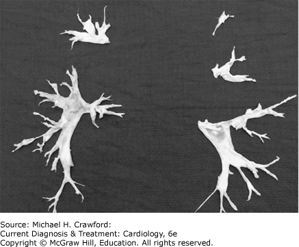
Chronic thromboembolic PH (CTEPH) occurs in up to 4% of patients following acute pulmonary embolism (PE) or 10% following recurrent PE. However, 50% of patients diagnosed with CTEPH have no antecedent history of a deep vein thrombosis or PE. Why some patients develop CTEPH is not well understood. Surgical specimens from patients undergoing pulmonary endarterectomy (PEA) demonstrate organization of thromboembolic material as well as vascular remodeling characterized by intimal thickening with deposition of collagen and hemosiderin, atherosclerosis, and calcification (Figure 30–3). Vasculopathy and pathologic remodeling of unobstructed downstream pulmonary arterioles like that which occurs in idiopathic PAH may also develop.
Figure 30-3. Surgical specimens obtained during pulmonary endarterectomy for the treatment of chronic thromboembolic pulmonary hypertension. (Reproduced with permission from Kim Kerr, MD, Professor of Medicine, UCSD.)
Excluding CTEPH as a cause of PH is important because mortality is high without appropriate surgical management. Excluding CTEPH as a cause of PH requires evaluation with a ventilation-perfusion scan followed by confirmatory conventional pulmonary angiography. Computed tomography (CT) pulmonary angiography is insensitive and does not exclude the diagnosis. If a diagnosis of CTEPH is suspected, early referral to a specialized center where PEA is performed is recommended. Balloon pulmonary angioplasty (BPA) may be of benefit in selected patients.
E. Pulmonary Hypertension with Unclear and Multifactorial Mechanism
This category of PH encompasses a diverse group of disorders that cause PH by mechanisms that are not easily characterized.
F. Right Ventricular Failure Resulting from Pulmonary Hypertension
PH increases the afterload of the right ventricle, resulting in compensatory right ventricle hypertrophic remodeling so that the right ventricle can maintain the CO. Early on, the right ventricle can have supernormal function and normal-to-reduced chamber dimensions. Chronic right ventricular pressure overload results in persistent upregulation of proproliferative neurohormones, endothelin-1, and cytokines. These mediators contribute to the development of maladaptive hypertrophy with fibrosis and diastolic dysfunction (Figures 30–4 and 30–5).
Figure 30-4. Appearance of the right ventricle in pulmonary hypertension. A: A transverse section through a normal heart illustrates a crescentic right ventricular cavity, thin right ventricular free wall (arrows), and round left ventricular cavity (line). B: A transverse section of a heart from a patient who had severe pulmonary hypertension showing dilation of the right ventricular cavity giving the right ventricle a more spherical shape than in the normal heart. Also seen is thickening of the right ventricular free wall (arrows) and flattening of the interventricular septum (line). LV, left ventricle; RV, right ventricle. (Reproduced, with permission, from Barnett CF, et al. Pulmonary hypertension due to lung disease. In: Broaddus VC, et al, eds. Murray and Nadels Textbook of Respiratory Medicine, 6th ed. Philadelphia: W.B. Saunders; 2016:10501065.e1055. Copyright © Elsevier.)
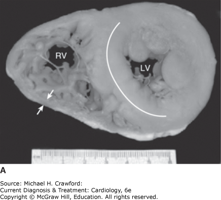
Figure 30-4.
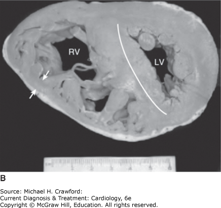
Figure 30-5. Pathophysiology of right ventricular dysfunction in pulmonary hypertension. AV-DO2, arteriovenous oxygen differential; CO, cardiac output; LV, left ventricular; RAP, right atrial pressure; RV, right ventricular.
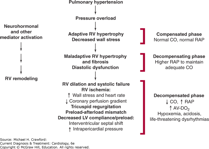
If exposure to elevated PAP continues, the right ventricle dilates. This is an ominous sign because it signifies the presence of increased right ventricular wall stress. Increased wall stress, when coupled with increased heart rate, results in increased myocardial oxygen demand. This develops in concert with a reduction in the epicardial-to-endocardial coronary perfusion gradient secondary to increased right ventricular end-diastolic pressure. In sum, these changes result in a myocardial oxygen supply-demand mismatch and right ventricular ischemia. Right ventricular ischemia worsens systolic function, increases end-diastolic pressure, and promotes further ventricular enlargement. Tricuspid regurgitation secondary to annular dilatation often occurs and serves to further reduce effective right ventricular forward output.
Right ventricular dilation, in the setting of an intact pericardium, results in a shift of the interventricular septum toward the left ventricle. This, especially when coupled with increased intrapericardial pressure, impedes left ventricular filling (preload). In turn, when left ventricular filling is impaired, systemic CO is reduced.
Multiple mechanisms contribute to the development of hypoxemia in PH, even when intrinsic pulmonary disease is absent. Pulmonary vascular remodeling and in situ thromboses disrupt normal capillary-alveolar gas exchange and raise the alveolar-to-arterial oxygen gradient. Increased pulmonary pressures can increase right atrial pressure and cause shunting through a patent foramen ovale.
HumbertM, GuignabertC, BonnetS,et al. Pathology and pathobiology of pulmonary hypertension: state of the art and research perspectives. Eur Respir J. Jan 2019;53(1):1801887. 30545970
HumbertM,et al. 2022 ESC/ERS Guidelines for the diagnosis and treatment of pulmonary hypertension. Eur Heart J. Aug 26 2022;36017548
HustonJH, ShahSJ. Understanding the pathobiology of pulmonary hypertension due to left heart disease. Circ Res. Apr 29 2022;130(9):13821403. 35482841
PapamatheakisDG, PochDS, FernandesTM, KerrKM, KimNH, FedulloPF. Chronic thromboembolic pulmonary hypertension: JACC Focus Seminar. J Am Coll Cardiol. Nov 3 2020;76(18):21552169. 33121723
SinghN, DorfmullerP, ShlobinOA, VentetuoloCE. Group 3 pulmonary hypertension: from bench to bedside. Circ Res. Apr 29 2022;130(9):14041422. 35482836
SouthgateL, MachadoRD, GrafS, MorrellNW. Molecular genetic framework underlying pulmonary arterial hypertension. Nat Rev Cardiol. Feb 2020;17(2):8595. 31406341
Vonk NoordegraafA, ChinKM, HaddadF,et al. Pathophysiology of the right ventricle and of the pulmonary circulation in pulmonary hypertension: an update. Eur Respir J. Jan 2019;53(1):1801900. 30545976