Tachyarrhythmias occur as a result of three main mechanisms: reentry, which is most common; enhanced or abnormal automaticity; and triggered activity.
Reentrant arrhythmias sustain themselves by repetitively following a revolving pathway comprising two limbs, one that takes the impulse away from and one that carries it back to the site of origin. For reentry to exist, an area of slow conduction must occur, and each limb must have a different refractory period (see the discussion on AV nodal reentrant tachycardia). In this situation, ectopic beats or pacing (by inducing refractoriness in one limb of the circuit) can initiate a reentrant tachycardia. Once established, ectopic beats or pacing can also terminate the tachycardia by interfering with impulse propagation in one of the limbs.
The second mechanism, automaticity, refers to spontaneous and, often, repetitive firing from a single focus, which may either be ectopic or may originate in the sinus node. This mechanism comprises two subcategories. Enhanced automaticity is defined as a focus that fires spontaneously and may originate in the sinus node, subsidiary pacemakers in the atrium including the Eustachian ridge, Bachmann bundle, coronary sinus and AV valves, the AV node, His-Purkinje system, and the ventricles. Abnormal automaticity is usually secondary to a disease process causing alterations in ionic flow that produces a less negative resting diastolic membrane potential. Threshold potential is therefore more easily attained, thereby increasing the probability of a sustained arrhythmia.
The third mechanism, triggered arrhythmias, depends on oscillations in the membrane potential that closely follow an action potential. In the absence of a new external electrical stimulus, these oscillations, or after-depolarizations, cause new action potentials to develop. Thus, each new action potential results from the previous action potential. These arrhythmias can be produced by early or late after-depolarization, depending on the timing of the first after-depolarization relative to the preceding action potential (the one that spawned the triggered activity). In early after-depolarizations, membrane repolarization is incomplete, which allows an action potential to be initiated by a subthreshold stimulus. This type is often associated with electrolyte disturbance and may be the mechanism responsible for arrhythmogenesis related to the prolonged QT syndrome and torsades de pointes caused by quinidine. With delayed after-depolarization, membrane repolarization is complete, but an abnormal intracellular calcium load causes spontaneous depolarization. The high calcium levels can be related to the inhibition of the sodium pump by drugs such as digoxin. In either type of arrhythmia, the process may be repetitive and lead to a sustained tachycardia.
General Diagnostic ApproachA systematic approach to interpreting the 12-lead electrocardiogram (ECG) will allow accurate determination of the type of SVT in most cases (Figure 11–1). The first step is to determine whether the rhythm is regular or irregular. If it is irregular, the rhythm is likely either atrial fibrillation, atrial flutter with variable conduction, or multifocal atrial tachycardia (MAT). The appearance of the P waves or lack of P waves will usually distinguish between these three entities. In atrial fibrillation, there is chaotic atrial activity. In atrial flutter, P waves are seen at a rate of 240–320 bpm. In MAT, there are P waves preceding each QRS complex, and there are at least three different P-wave morphologies.
Figure 11-1. Algorithm for distinguishing supraventricular tachycardias. AF, atrial fibrillation; AFL, atrial flutter; AT, atrial tachycardia; AVNRT, atrioventricular nodal reentrant tachycardia; AVRT, atrioventricular reciprocating tachycardia; JT, junctional tachycardia; MAT, multifocal atrial tachycardia; SN, sinus node; ST, sinus tachycardia; SVT, supraventricular tachycardia.
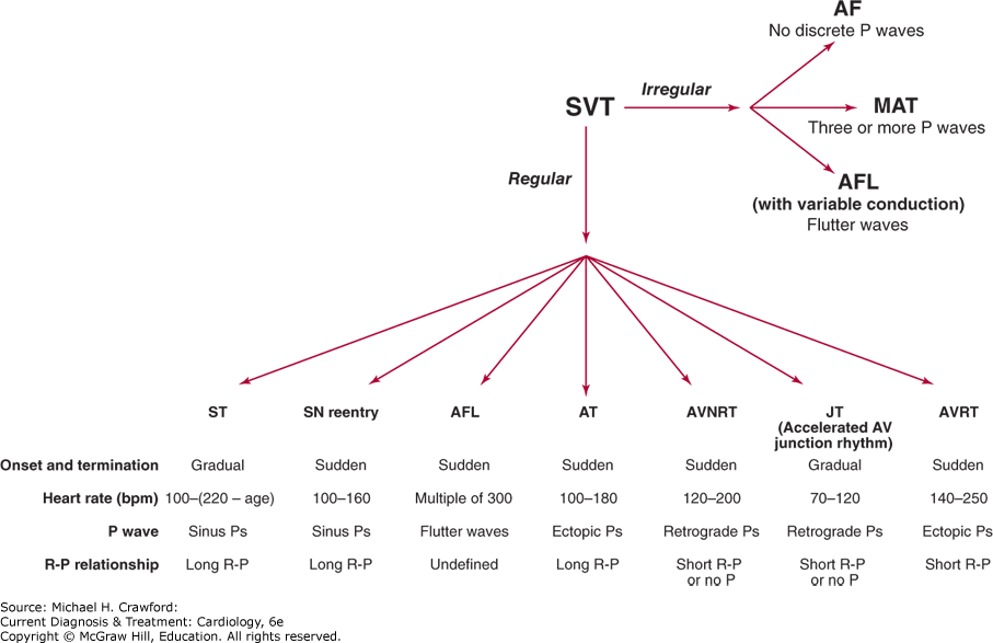
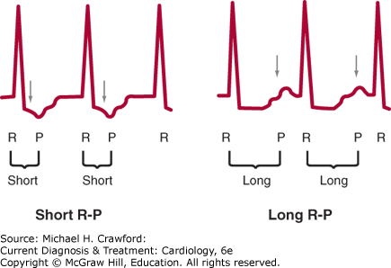
If the SVT is regular, there are several main types of SVT to consider. The SVT could be sinus tachycardia, sinus node reentry, atrial flutter, atrial tachycardia, AV nodal reentrant tachycardia (AVNRT), junctional tachycardia, or atrioventricular reciprocating tachycardia (AVRT). The type of regular SVT can be usually identified by examining four aspects of the 12-lead ECG: onset and termination, heart rate, P-wave morphology, and R-P relationship. Sinus tachycardia and junctional tachycardia typically have very gradual onset, whereas the other SVTs usually start and stop more suddenly. Rate can also be helpful since sinus tachycardia cannot typically go over 220 bpm minus age, and the heart rate in atrial flutter is often a multiple of 300. P-wave morphology can be helpful because retrograde P waves (negative in the inferior leads: II, III, and aVF) favor AVNRT and junctional tachycardia. Finally, R-P relationship refers to the distance from the R wave to the next P wave during tachycardia. If this distance is longer than the P-R interval, the SVT is termed “long R-P,” whereas if this distance is short, it is termed “short R-P” (Figure 11–2).
Figure 11-2. Short R-P refers to a regular supraventricular tachycardia (SVT) where the R-P interval is shorter than the P-R interval. Long R-P refers to a regular SVT where the R-P interval is longer than the P-R interval.