Patients usually have a history of murmur during infancy, and cyanosis occurs later in childhood. Exertional dyspnea is the most commonly encountered symptom. Chest pain, hemoptysis, and presyncope are less common. Transient bacteremia can result in brain abscess as a result of right-to-left shunting and entry of bacteria into the cerebral circulation without the normal filtering through the pulmonary circulation.
Physical examination reveals cyanosis (involving the legs when the cause is PDA); cardiovascular examination is most remarkable for findings associated with pulmonary hypertension. The LV impulse is not displaced and an RV parasternal heave is present. The jugular venous pressure may be elevated in the presence of RV failure, and the a wave may be prominent. The first heart sound is normal, and P2 is markedly accentuated. A systolic murmur of tricuspid regurgitation may be present, and a high-pitched diastolic murmur of pulmonary regurgitation (Graham Steell murmur) is common. In the presence of RV failure, hepatomegaly, ascites, and peripheral edema may be present.
1. Electrocardiography and Chest Radiography
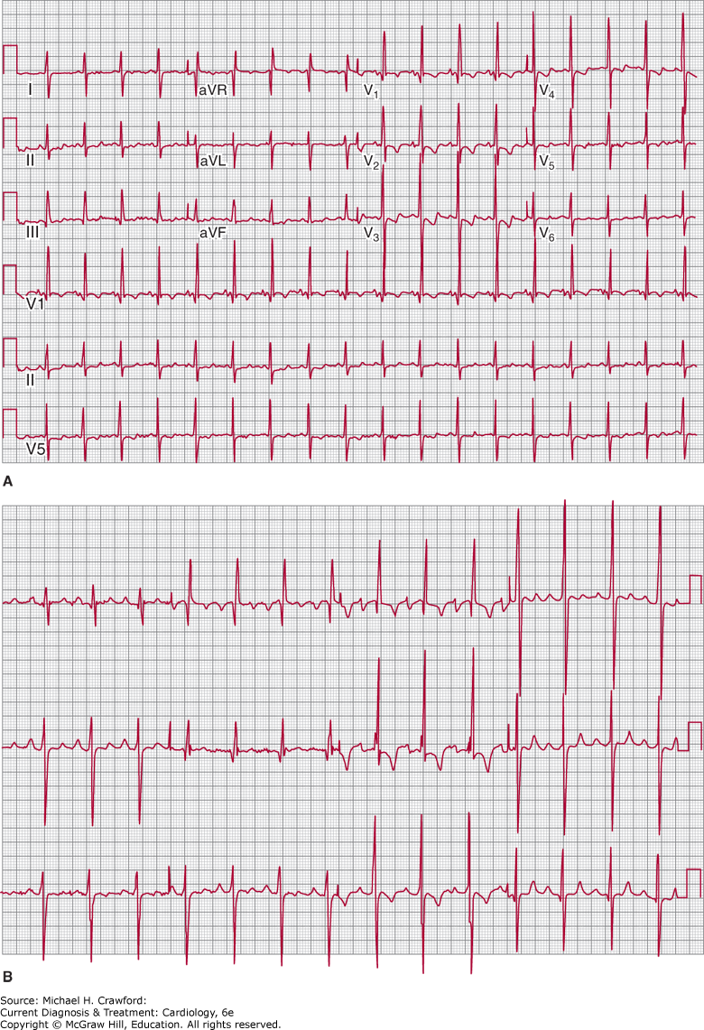
The ECG shows evidence of right atrial enlargement and RVH with a rightward axis (Figure 31–26A). The presence of a leftward or superior axis suggests an ostium primum ASD or AV canal defect as the underlying cause (Figure 31–26B). Chest radiograph findings include RV enlargement with filling in of the retrosternal air space, prominent proximal pulmonary arteries with pulmonary oligemia, and pruning of the peripheral pulmonary vessels.
Figure 31-26. A: Electrocardiograph in Eisenmenger syndrome from ostium secundum atrial septal defect with right ventricular hypertrophy and right-axis deviation. B: Electrocardiograph in Eisenmenger syndrome and atrioventricular canal defect with right ventricular hypertrophy and left anterior hemiblock.
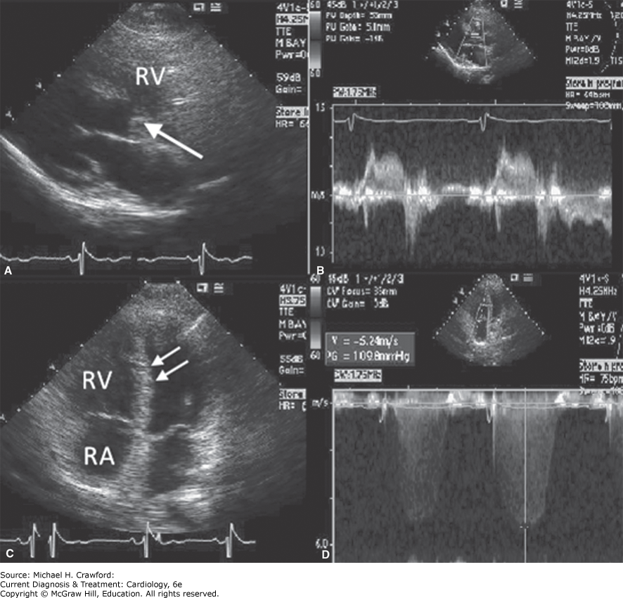
Severe RVH and right atrial enlargement are evident (Figure 31–27A). RV function may be normal until the late stages of the disease, at which time the right atrium enlarges. The LV appears small and underfilled; the septum deviates toward the LV. The level of shunt can be determined by two-dimensional imaging, aided by color-flow Doppler and saline contrast injection (Figure 31–27B). When a VSD is present, the flow velocity across the defect is low because of pressure equalization between the two ventricles. On the other hand, the tricuspid regurgitant velocity is increased and can be used to estimate the peak RV systolic pressure (Figure 31–27C). Pulmonary insufficiency with a high-velocity regurgitant jet is a frequent finding. Other valvular lesions are uncommon except in ostium primum ASD or AV canal defects, when mitral regurgitation is commonly present.
Figure 31-27. A: Transthoracic echocardiogram in a 29-year-old patient with a large perimembranous ventricular septal defect (arrow). B: With bidirectional shunting. C: There is bowing of the septum to the left (double arrows), a dilated right atrium (RA), and dilated and hypertrophied right ventricle (RV). D: Systemic right ventricular pressures were estimated from a continuous wave Doppler tracing of the tricuspid regurgitation jet. Pressure gradient (PG) 109.8 mmHg plus RA pressure = RV pressure.
Eisenmenger syndrome can usually be differentiated from primary pulmonary hypertension noninvasively by TTE, although shunting across a patent foramen ovale may mimic an ASD with Eisenmenger syndrome and a PDA may be missed on color-flow Doppler in the presence of severe pulmonary hypertension. In these cases, further investigation with TEE or cardiac catheterization and sometimes cardiac MRI/MRA may be indicated.
The pathognomonic hemodynamic findings are elevated pulmonary artery pressure, increased PVR, and right-to-left shunting. The degree of residual left-to-right shunt should be measured. Oxygen should be administered during catheterization to determine whether pulmonary vascular reactivity persists. If PVR falls during oxygen or nitric oxide administration, increased left-to-right shunting can be measured. In this case, the patient may be a candidate for pulmonary vasodilator therapy and surgical repair.
The arterial oxygen saturation by pulse oximetry or arterial blood gas measurements is markedly decreased. The hematocrit is elevated, with an overall increase in red cell mass. Iron deficiency is common, particularly after injudicious phlebotomies. Hyperuricemia caused by increased red cell turnover may be present.