Introduction ⬇
| Essentials of Diagnosis Pulmonary hypertension A loud pulmonic valve closure sound (P2), a right-sided S4, or a right ventricular heave. Electrocardiographic evidence of right ventricular hypertrophy. Presence of sustained elevation in mean pulmonary artery pressure ≥ 20 mm Hg.
Pulmonary arterial hypertension A subset of pulmonary hypertension. Elevated mean pulmonary artery pressure ≥ 20 mm Hg and pulmonary arterial wedge pressure ≤ 15 mm Hg and PVR > 2 Wood units. Appropriate clinical context.
|
Pulmonary hypertension (PH) describes the finding of a mean pulmonary artery pressure (mPAP) ≥ 20 mm Hg and may occur in many settings. In most cases, PH results from a left heart disease (LHD) that increases pulmonary artery pressure (PAP) by transmission of elevated left heart filling pressures or from lung disease causing hypoxia-mediated pulmonary vasoconstriction. The term pulmonary arterial hypertension (PAH) describes a specific group of diseases characterized hemodynamically by an mPAP ≥ 20 mm Hg and normal left heart filling pressures (pulmonary artery wedge pressure [PAWP] ≤ 15 mm Hg) resulting from vasoconstriction and arteriopathy of the precapillary pulmonary arterioles. PAH may be idiopathic or associated with one or more underlying diseases such as connective tissue disease, human immunodeficiency virus (HIV) infection, or portal hypertension. In patients clinically suspected of having PH, echocardiography is often the first test performed to estimate PAP and evaluate right ventricular function. Further comprehensive testing is required to make a diagnosis and establish the etiology of PH.
PH commonly occurs in patients with LHD and hypoxia from lung disease. The development of PH in patients with heart or lung disease is an ominous sign and is generally associated with reduced survival. Treatment in such cases is directed at the underlying condition (ie, bronchodilators, supplemental oxygen, valve repair, or heart failure treatment). The use of PAH-specific therapy in this setting is generally not beneficial and can worsen symptoms and mortality. In patients with PAH, vasoconstriction and pulmonary vascular arteriopathy cause increased pulmonary vascular resistance (PVR) and increased PAP, ultimately causing right heart failure and death. In this setting, PAH-specific therapy improves symptoms, exercise tolerance, and survival. Selection of PAH therapies is complex, and the prescriber must consider PAH severity, associated diseases, toxicities, and drug interactions and provide close follow-up. Early referral and collaboration with a PH specialty center are recommended for confirmation of diagnosis, initiation of appropriate therapy, and monitoring of response to treatment. Despite significant advances in understanding of the clinical profile, pathobiology, and treatment of PAH, delays in diagnosis remain common and patient outcomes remain poor.
General Considerations ⬆ ⬇
PH is defined as a sustained elevation in the mPAP of ≥ 20 mm Hg at rest. This contrasts with a normal mPAP of 12–16 mm Hg. In healthy humans, the small arteriovenous pressure gradient generated by blood flow across the pulmonary circulation results from the large total vascular surface area and high pulmonary vascular compliance. The PVR is quantified using Ohm’s law (PVR = [mPAP – PAWP]/cardiac output [CO]) and describes the relationship between the pressure gradient and blood flow.
Clinical Findings ⬆ ⬇
A. Symptoms & Signs
PAH frequently presents with nonspecific symptoms such as dyspnea on exertion, fatigue, or chest pain. These symptoms are often attributed to other diseases such as asthma, anxiety, or, if present, connective tissue disease or HIV. Exertional presyncope or syncope, edema, and ascites develop in more advanced right heart failure. A modified New York Heart Association (NYHA) classification system is used to describe patients with PH (Table 30–2).
Table 30–2. New York Heart Association (NYHA) and World Health Organization (WHO) Functional Classes in Pulmonary Hypertension
| Class | Symptoms/Function |
|---|
| NYHA I/WHO I | No limitation in physical activity. Ordinary physical activity does not cause undue dyspnea, fatigue, chest pain, or near-syncope. |
| NYHA II/WHO II | Slight limitation in physical activity. Ordinary physical activity causes undue dyspnea, fatigue, chest pain, or near-syncope. |
| NYHA III/WHO III | Marked limitation in physical activity. Less than ordinary physical activity causes undue dyspnea, fatigue, chest pain, or near-syncope. |
| NYHA IV/WHO IV | Inability to carry out any physical activity without symptoms. Patients manifest signs of right heart failure. Dyspnea and/or fatigue may be present at rest. |
B. Physical Examination
Physical examination of the patient with PH can reveal clues not only about the presence or severity of PH but also about the underlying cause. Classic examination findings that occur late with markedly elevated PAP include a loud pulmonic valve closure (P2) or an early systolic ejection click. A right-sided S3 or S4 gallop is heard in patients with significant elevation in the right ventricular end-diastolic pressure. With right ventricular hypertrophy and enlargement, a left parasternal lift can be palpated.
Often the holosystolic murmur of tricuspid regurgitation (although frequently without the classically described respiratory variation) and the less common diastolic murmur of pulmonic insufficiency are noted in patients with PH. Cool extremities, diminished peripheral pulses, jugular venous distention, peripheral edema, and ascites develop with progressive right heart failure.
A murmur of mitral or aortic stenosis or a left-sided S3 or S4 gallop suggests left-sided heart disease, whereas wheezing and diminished breath sounds may be clues to the presence of pulmonary parenchymal disease. Jaundice and spider angiomata may point to the presence of cirrhosis and portal hypertension. Connective tissue diseases may present not only with signs of PH but also with Raynaud phenomenon, arthritis, rashes, or other skin changes such as sclerodactyly.
C. Diagnostic Studies
1. Electrocardiography
The electrocardiogram (ECG) in patients with significant PH typically shows right ventricular hypertrophy. Classic ECG findings include right axis deviation and right atrial enlargement. Incomplete or complete right bundle branch block is also common. The ECG criteria for right ventricular hypertrophy and a typical ECG in a patient with PH are shown in Table 30–3 and Figure 30–6, respectively. However, the ECG is not sensitive enough to exclude a diagnosis of PH.
Figure 30-6. Electrocardiogram in a patient with pulmonary arterial hypertension and right ventricular hypertrophy.
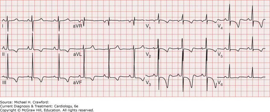
Table 30–3. Electrocardiographic Criteria for Right Ventricular Hypertrophy
Right axis deviation (axis > +90 degrees) R/S amplitude ratio in lead V1> 1.0 mm R-wave amplitude in V1≥ 7 mm S-wave amplitude in V1< 2 mm qR pattern in precordial lead V1 R-wave amplitude in lead V1 + S-wave amplitude in V5 or V6> 10.5 mm R/S amplitude ratio in V5 or V6< 1.0 mm |
2. Chest Radiography
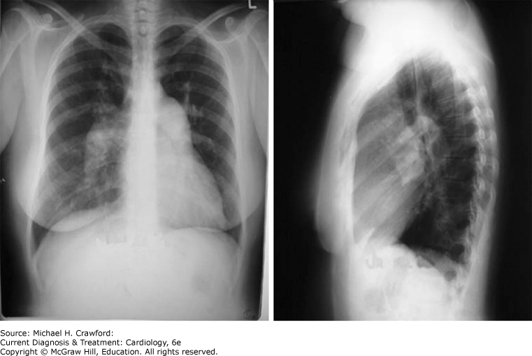
The chest radiograph in a patient with PAH may show enlargement of the main pulmonary artery and its major branches with a reduction in the number of distal vessels referred to as pruning. A lateral image will show filling of the retrosternal space, which signifies right ventricular enlargement. Pulmonary venous congestion and left atrial or left ventricular enlargement suggest the presence of a left-sided cause of PH. Hyperinflated lung fields or bullous changes point to PH from lung disease. A classic chest radiograph of a patient with PAH is shown in Figure 30–7.
Figure 30-7. Chest radiograph in pulmonary arterial hypertension demonstrating enlargement of the central pulmonary arteries with peripheral pruning of the pulmonary vasculature. Also notable is the reduction in the retrosternal air space on the lateral view and the distinct lack of pulmonary pathology.
3. Echocardiography
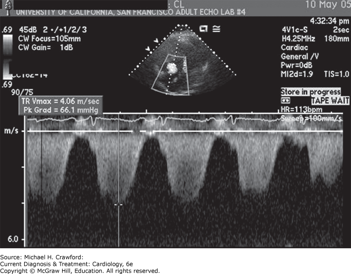
Echocardiography is often the first test ordered in a patient with suspected PH and may reveal direct or indirect evidence of elevated PAP. Echocardiography is used to estimate the pulmonary artery systolic pressure (PASP). The peak velocity (v) of the tricuspid regurgitant (TR) jet is determined from the continuous wave spectral Doppler signal and entered into the modified Bernoulli equation along with the right atrial pressure (RAP), estimated by evaluating respiratory change in the inferior vena cava diameter: PASP = 4v2 + RAP (Figure 30–8). Echocardiographic estimates of PASP are often inaccurate, especially if the TR jet is minimal or eccentric. Therefore, a low estimated PASP does not exclude PH in patients in whom PH is clinically suspected.
Figure 30-8. A continuous wave Doppler recording of the tricuspid regurgitation velocity. The pulmonary artery systolic pressure is calculated by entering the peak tricuspid velocity into the modified Bernoulli equation and adding the right atrial pressure (PASP = 4v2 + RAP). PASP, pulmonary artery systolic pressure; v, peak tricuspid regurgitant velocity; RAP, right atrial pressure.
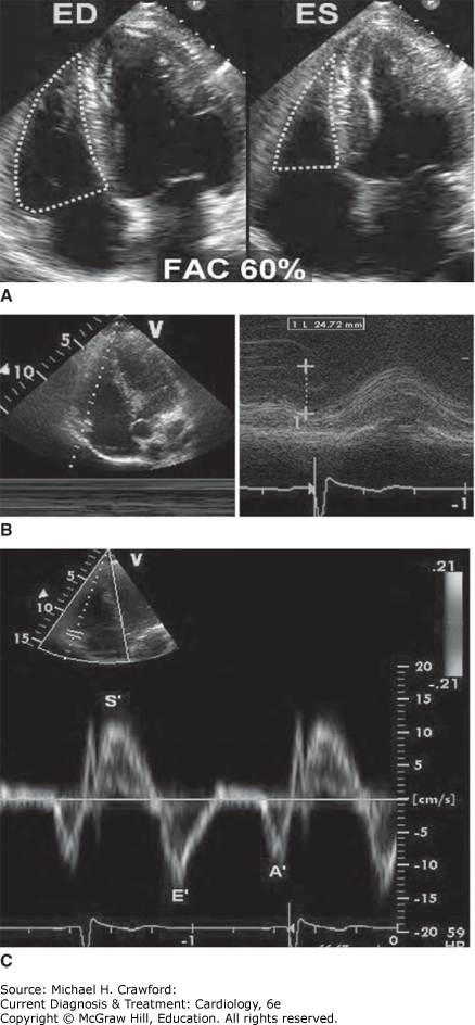
Structural changes seen on echocardiography may suggest that PH is present. Over time, elevated PAP results in right atrial or right ventricular enlargement, right ventricular hypertrophy, and pulmonary artery enlargement. Ventricular interdependence results in flattening or leftward shift of the interventricular septum and, if seen during systole, suggests pressure overload or, if seen during diastole, suggests volume overload of the right ventricle. The left ventricle is often small and underfilled in PH, with normal systolic function (Figure 30–9). Accurate assessment of the right ventricular systolic function in patients with PH is important because impaired function is a predictor of poor survival. Approaches to right ventricular function assessment are described; however, they are subject to multiple technical limitations so that the opinion of an experienced echocardiographer is necessary (Figure 30–10).
Figure 30-9. Transthoracic echocardiogram of a healthy patient compared to a patient with pulmonary arterial hypertension (PAH). A: Apical four-chamber view of a normal heart. B: Apical four-chamber view of the heart from a patient with PAH demonstrating enlargement of the right atrium and right ventricle (RV). C: Parasternal short-axis view of a normal heart. D: Parasternal short-axis view demonstrates RV enlargement, a small left ventricular (LV) cavity, and leftward shifting (flattening) of the interventricular septum. (Reproduced with permission from Barnett CF, et al. Pulmonary hypertension due to lung disease. In: Broaddus VC, et al, eds. Murray and Nadels Textbook of Respiratory Medicine, 6th ed. Philadelphia: W.B. Saunders; 2016:10501065.e1055. Copyright © Elsevier.)
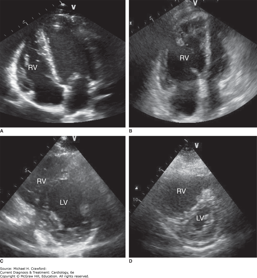
Figure 30-10. Approach to assessment of right ventricular function in patients with pulmonary hypertension. A: Right ventricular fractional area change (FAC). B: Tricuspid annular plane systolic excursion C: Tissue Doppler of tricuspid annular systolic velocity (S'). ED, end diastolic; ES, end systolic; E', tricuspid annular plane early diastolic; A', late diastolic velocities.
The careful review of the echocardiogram is required to exclude LHD as a cause of PH. HFrEF and valvular heart disease are generally easily diagnosed on a routine echocardiogram. Diagnosing HFpEF is more challenging and should be suspected when PH is seen in a patient with left atria or left ventricular enlargement or left ventricular hypertrophy. Additional maneuvers, such as injection of agitated saline, should be performed to identify cardiac and intrapulmonary shunts.
4. Ventilation-Perfusion Lung Scintigraphy
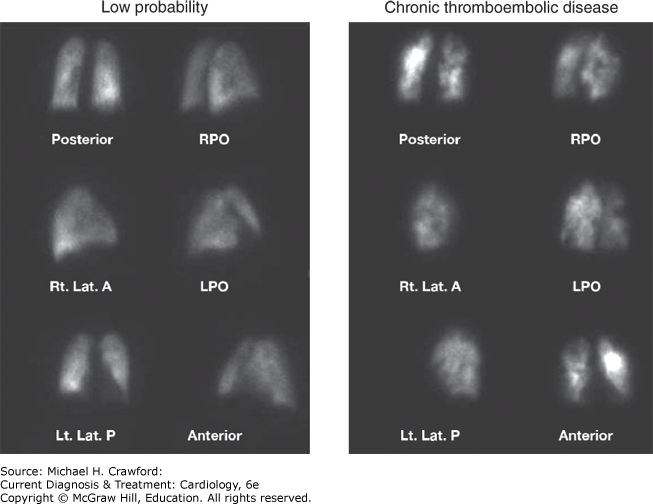
Ventilation-perfusion (V/Q) lung scintigraphy is required to exclude CTEPH as the etiology of PH because CT pulmonary angiography is insensitive to this diagnosis. In PAH, the V/Q scan may reveal a normal perfusion pattern, or it may show diffuse, patchy perfusion defects. Parenchymal lung disease can also result in perfusion scan abnormalities, but typically, these are matched by ventilatory defects. In CTEPH, the lung perfusion scan demonstrates one or more segmental defects mismatched by the ventilation scan (Figure 30–11). An abnormal V/Q scan should prompt referral to a CTEPH center and conventional pulmonary angiography to confirm the diagnosis of CTEPH and consideration for surgical PEA.
Figure 30-11. Perfusion lung scans from a normal patient and a patient with chronic thromboembolic disease. The scan on the left shows uniform perfusion. The scan on the right shows multiple perfusion defects consistent with the diagnosis of chronic thromboembolic disease. LPO, left posterior oblique; RPO, right posterior oblique.
5. Pulmonary Function Testing
Pulmonary function testing is helpful in PH because it can establish the diagnosis of underlying obstructive or restrictive pulmonary disease. Interpretation of pulmonary function test results should be tempered by an awareness that PAH can reduce diffusing capacity of carbon monoxide. If the total lung capacity is less than 70% of predicted, a high-resolution CT scan should be considered to evaluate for ILD.
6. Computed Tomography and Magnetic Resonance Imaging
Chest CT or magnetic resonance imaging (MRI) scans may reveal other causes of PH such as fibrosing mediastinitis and cystic fibrosis, as well as infiltrative or granulomatous lung diseases. Findings on CT pulmonary angiography may suggest a diagnosis of CTEPH; however, the test is not sensitive enough to exclude the diagnosis of CTEPH.
7. Cardiac Catheterization and Pulmonary Angiography
RHC represents the gold standard test to establish the diagnosis of PH, ascertain its etiology, establish severity and prognosis, evaluate vasoreactivity, and guide therapy. RHC should be performed by a clinician with experience in the evaluation and management of patients with PH.
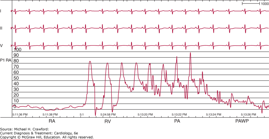
During RHC, hemodynamic measurements should include right atrial pressure, right ventricular pressure, PAP, PAWP, and the CO (Figure 30–12). The PVR is calculated using the following equation (mPAP – PAWP)/CO. Arterial and venous oxygen saturations obtained during RHC are used to detect and determine the presence and severity of shunts. An elevated PAWP suggests LHD, such as aortic or mitral valve disease, HFrEF, or HFpEF as the cause of PH. In elderly patients with diabetes, hypertension, left ventricular hypertrophy, or left atrial enlargement, administration of a fluid bolus or exercise during RHC should be considered to exclude the presence of otherwise unrecognized HFpEF.
Figure 30-12. Hemodynamic tracing from a patient with pulmonary hypertension. Waveforms that are obtained as the catheter passes from the right atrium (RA) through the right ventricle (RV), pulmonary artery (PA), and into the pulmonary artery wedge position. Notable findings include the markedly elevated pulmonary artery pressure and the normal pulmonary artery wedge pressure (PAWP).
Vasoreactivity testing during RHC is used to identify the small proportion of patients with idiopathic or heritable PAH who can be successfully treated with calcium channel blockers and is not useful in patients with PAH from other underlying causes. Acute vasoreactivity testing may result in hemodynamic instability and should be performed with an RHC in place in a monitored setting with the supervision of experienced physicians. Vasodilator testing is usually performed by administering inhaled nitric oxide at a dose of 20 parts per million for 10 minutes. A reduction in mPAP greater than 10 mm Hg with a final mPAP ≤ 40 mm Hg without a reduction in the CO or increase in the PAWP identifies patients who can be treated with calcium channel blockers.
Although not routinely performed, pulmonary angiography is useful to confirm the diagnosis of CTEPH and to determine if surgical PEA can be performed.
8. Lung Biopsy
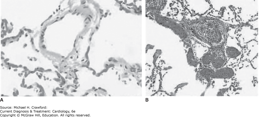
Lung biopsy rarely provides useful diagnostic information and is associated with significant risk in patients with PH. When performed, lung biopsy may reveal findings such as injected particulate matter in injection drug users, arterialization of pulmonary venules in pulmonary veno-occlusive disease, or typical findings of ILD. Typical pathologic findings of PAH are illustrated in Figure 30–13.
Figure 30-13. Pathologic appearance of a normal pulmonary arteriole compared to a pulmonary arteriole in pulmonary arterial hypertension. A: Normal pulmonary arteriole. B: Pulmonary arteriole from a patient with pulmonary arteriole hypertension demonstrating a vascular endothelial cell proliferation and smooth muscle cell hypertrophy and a plexiform lesion. (Reprinted, with permission, from Barnett CF, et al. Pulmonary hypertension due to lung disease. In: Broaddus VC, et al, eds. Murray and Nadels Textbook of Respiratory Medicine, 6th ed. Philadelphia: W.B. Saunders; 2016:10501065.e1055. Copyright © Elsevier.)
9. Functional Capacity and Exercise Testing
Determination of each patient’s World Health Organization functional classification and symptom-limited exercise testing should be performed during the evaluation of all patients with PH. Exercise testing, particularly the 6-minute walk (6MW) test, has been shown to predict mortality and allows for objective assessment of symptom burden. Exercise tolerance should be assessed prior to initiation of therapy with the 6MW test, which is then repeated often to monitor response to treatment.
10. Other Studies
Overnight pulse oximetry should be performed to identify patients who require supplemental oxygen to treat nocturnal hypoxemia. If sleep-disordered breathing is suspected, a full sleep study should be performed. Testing of liver enzymes and liver ultrasound is used to look for evidence of liver dysfunction and portal hypertension. Screening for connective tissue diseases including scleroderma, rheumatoid arthritis, systemic lupus erythematosus, mixed connective tissue disease, and polymyositis should be performed with appropriate serologic and immunogenetic studies. Thyroid function and HIV testing should also be performed in all patients. Schistosomiasis can cause PAH and should be excluded in patients from endemic areas with PAH.
FarrellC, BalasubramanianA, HaysAG,et al. A clinical approach to multimodality imaging in pulmonary hypertension. Front Cardiovasc Med. 2021;8:794706. 35118142
MaronBA, Kovacs,Vaidya A,et al. Cardiopulmonary hemodynamics in pulmonary hypertension and heart failure: JACC Review Topic of the Week. J Am Coll Cardiol. 2020 Dec 1;76(22):26712681. 33243385
Remy-JardinM, RyersonCJ, SchieblerML,et al. Imaging of pulmonary hypertension in adults: a position paper from the Fleischner Society. Eur Respir J. Jan 2021;57(1):2004455. 33402372
RuoppNF, CockrillBA. Diagnosis and treatment of pulmonary arterial hypertension: a review. JAMA. Apr 12 2022;327(14):13791391. 35412560
Differential Diagnosis ⬆ ⬇
Determining if a patient has PAH versus PH from heart disease, lung disease, or CTEPH is critically important due to significant differences in management and survival (see Table 30–1). For example, endothelin receptor antagonists significantly improve outcomes in patients with PAH, whereas, by contrast, they worsen symptoms and increase hospitalization in patients with LHD and ILD. A comprehensive diagnostic evaluation that integrates findings from the patient history and physical examination as well as the results of all diagnostic tests is mandatory so that underlying diseases can be correctly identified and treated and so that optimal therapy for PAH, if present, can be chosen. The diagnostic approach currently recommended in PH is shown in Figure 30–14.
Figure 30-14. Diagnostic algorithm for pulmonary arterial hypertension updated at the Fifth World Symposium on Pulmonary Hypertension. BGA, blood gas analysis; CHD, congenital heart disease; CTD, connective tissue disease; CTEPH, chronic thromboembolic pulmonary hypertension; DLCO, diffusion capacity of the lung for carbon monoxide; ECG, electrocardiogram; HR-CT, high-resolution computed tomography; PA, pulmonary angiography; PAH, pulmonary arterial hypertension; PAPm, mean pulmonary artery pressure; PAWP, pulmonary arterial wedge pressure; PCH, pulmonary capillary hemangiomatosis; PEA, pulmonary endarterectomy; PFT, pulmonary function testing; PH, pulmonary hypertension; PVOD, pulmonary veno-occlusive disease; PVR, pulmonary vascular resistance; RHC, right heart catheter; RV, right ventricle; V/Q, ventilation/perfusion; WU, Woods units; x-ray, chest radiograph. (Reproduced with permission from Hoeper MM, et al. Definitions and diagnosis of pulmonary hypertension. J Am Coll Cardiol. 2013;62[25 Suppl]:D42D50.)

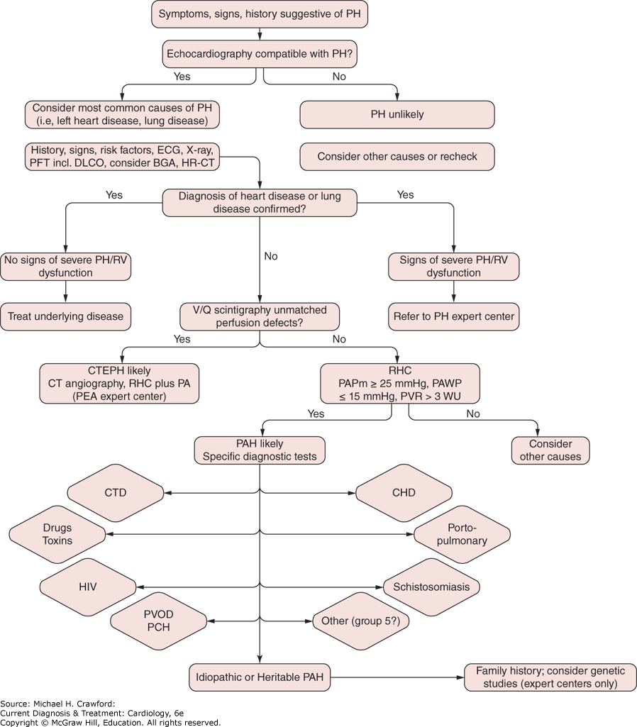
In many cases, an elevated PAP is first noted on echocardiography that is performed to evaluate symptoms of exertional dyspnea, edema, chest pain, presyncope, or syncope. LHD is the most common cause of PH, and often, HFrEF or severe valvular heart disease will be evident on the initial echocardiogram so that no further evaluation will be required.
Pulmonary disease is the second most common cause of PH and may be suggested by findings on chest radiograph, arterial oxygen saturation measurement, pulmonary function testing, sleep study, and chest CT scan.
A V/Q scan should be performed, and diagnosis of CTEPH should be considered in all patients with PH because it may coexist with PH from heart or lung disease.
A diagnosis of PAH is typically reached when other more common causes of PH have been excluded. Patients then undergo evaluation for secondary causes of PAH by performing serologic testing for connective tissue diseases, thyroid disease, and HIV. Imaging studies should be reviewed to exclude evidence of portal hypertension.
RHC is a required test in all patients in whom a diagnosis of PAH is being considered to confirm the diagnosis as well as determine prognosis and select initial therapy. RHC is also sometimes required to exclude a diagnosis of HFpEF. Reaching a final diagnosis requires integrating features of the clinical presentation together with hemodynamic findings and results of the entire diagnostic evaluation.
Treatment ⬆ ⬇
A. Pulmonary Arterial Hypertension
Improvement in exercise capacity, functional class, and survival are the goals of PAH treatment. Physical activity should be limited below levels that provoke symptoms. However, low-intensity physical activity or regular exercise to maintain skeletal muscle conditioning and overall cardiovascular fitness is beneficial. Supplemental oxygen should be administered to all patients to maintain an oxygen saturation more than 92% with exertion, as well as for travel to high altitudes and for air travel. Immunization against influenza and pneumococcus is recommended. Oral anticoagulation is no longer recommended in all forms of PAH. Anticoagulation can be considered in patients with idiopathic, heritable PAH, and drug-induced PAH on a case-by-case basis after an individualized risk-benefit analysis. Treatment is generally with warfarin adjusted to an international normalized ratio (INR) of 1.5–2.5.
Pregnancy in patients with PAH is associated with high mortality, so women of childbearing potential should be counseled to avoid pregnancy and should be prescribed appropriate contraception. Hemoglobin levels should be monitored regularly. In patients with Eisenmenger syndrome, erythrocytosis should be treated with phlebotomy if symptoms of hyperviscosity develop. Patients with PAH should not smoke. Sodium restriction is often necessary to minimize fluid retention.
Diuretic therapy is necessary for most patients to maintain euvolemia. Furosemide, even at high doses, may be inadequate due to poor absorption and variable pharmacodynamics. Alternative loop diuretics such as bumetanide and torsemide and combination with thiazide diuretics are often necessary. Atrial arrhythmias are best treated with digoxin, which is tolerated better than calcium channel blockers or β-blockers.
In patients with PAH and decompensated right heart failure, strategies to improve right ventricular function and restore end-organ perfusion include (1) afterload reduction with pulmonary vasodilators, such as oxygen, inhaled nitric oxide, prostanoids, endothelin receptor antagonists, and phosphodiesterase inhibitors; (2) preload reduction with diuretics; and (3) augmentation of right ventricular function with intravenous inotropic drugs such as dobutamine or milrinone.
A small percentage of patients (< 10%) with idiopathic or heritable PAH can be successfully treated with high-dose calcium channel blocker therapy. These patients can be identified by a reduction in mPAP of at least 10 mm Hg to ≤ 40 mm Hg without a reduction in CO or an increase in PAWP. In patients who do not meet all these criteria, calcium channel blocker therapy is not beneficial and should not be used to treat PAH.
Multiple therapies approved by the US Food and Drug Administration (FDA) to treat PAH are available. These agents target the endothelin, nitric oxide, and prostacyclin pathways. When used in the appropriate patient, these pulmonary vasodilators can improve symptoms, exercise capacity, and mortality (Table 30–4).
Table 30–4. U.S. Food and Drug Administration–Approved Drugs for the Treatment of Pulmonary Hypertension
| Drug | Study Characteristics | Positive Results | Disadvantages |
|---|
| Endothelin Receptor Antagonists (ERAs) |
Bosentan (A and B) | Name: BREATHE-1 Design: Double-blind Number: 213 | Improved 6-minute walk distance Improved dyspnea Delayed clinical worsening | Hepatic toxicity Teratogenic Fluid retention, peripheral edema, anemia, nasal congestion, sinusitis, flushing Monthly transaminase monitoring required |
Ambrisentan (A) | Name: ARIES-1 Name: ARIES-2 Design: Double-blind Number: 202 and 192, respectively | Improved 6-minute walk distance Delayed clinical worsening Improved hemodynamics Transaminase monitoring not required | Teratogenic Fluid retention, peripheral edema, anemia, nasal congestion, sinusitis, flushing |
Macitentan (A and B) | Name: SERAPHIN Design: Double-blind Number: 742 | Reduced incidence of composite end point of death, atrial septostomy, lung transplantation, IV or SC prostanoid therapy, or worsening PAH | Teratogenic Headache, nasopharyngitis, anemia |
Phosphodiesterase-5 (PDE-5) Inhibitors |
Sildenafil | Name: SUPER-1 Design: Double-blind Number: 278 | Improved 6-minute walk distance Improved dyspnea Improved hemodynamics | No delay in clinical worsening end point Headache, flushing, dyspepsia, epistaxis, visual disturbance Interactions with protease inhibitors |
Tadalafil | Name: PHIRST Design: Double-blind Number: 405 | Improved 6-minute walk distance Improved time to clinical worsening Improved hemodynamics Improved quality of life | Headache, myalgias, flushing, dyspepsia, epistaxis, visual disturbance |
Soluble guanylate cyclase agonists |
Riociguat | Name: PATENT-1 and -2 Design: Double-blind Number: 443 | Improved 6-minute walk distance Improved hemodynamics Improved time to clinical worsening Improved quality of life Reduced brain natriuretic peptide Improved WHO class Improved dyspnea | Teratogenic Headache, dyspepsia, edema, dyspepsia, nausea, dizziness Severe hypotension with PDE-5 inhibitors |
Riociguat | Name: CHEST-1 and -2 (Group 4 PH-CTEPH) Design: Double-blind Number: 261 | Improved 6-minute walk distance Improved hemodynamics Reduced brain natriuretic peptide Improved WHO class | Teratogenic Severe hypotension with PDE-5 inhibitors |
Prostanoids |
Epoprostenol, intravenous | Design: Open-label Number: 81 | Improved 6-minute walk distance Improved dyspnea Improved hemodynamics Improved survival | Indwelling central line Pump malfunction Flushing, jaw pain, thrombocytopenia, headache, dizziness, nausea/vomiting/diarrhea, abdominal pain, hypotension, rash |
Treprostinil, intravenous or subcutaneous | Design: Double-blind Number: 470 | Improved 6-minute walk distance Improved dyspnea Improved hemodynamics | Indwelling central line or subcutaneous catheter Pain, erythema at infusion site (subcutaneous) Flushing, jaw pain, thrombocytopenia, headache, dizziness, nausea/vomiting/diarrhea, abdominal pain, hypotension, rash |
Treprostinil, inhaled | Name: TRIUMPH Design: Double-blind Number: 470 | Improved 6-minute walk distance Improved quality of life Administration 4 times daily | No delay in clinical worsening or dyspnea No change in functional class Cough, headache, nausea, dizziness, flushing, throat irritation or pain |
Treprostinil, inhaled | Name: INCREASE (Group 3 PH-ILD) Design: Double-blind Number: 326 | Improved 6-minute walk distance Improved time to clinical worsening Reduced brain natriuretic peptide | Cough, headache, dyspnea, dizziness, nausea, fatigue, and diarrhea |
Treprostinil, oral | Name: FREEDOM-M Design: Double-blind Number: 349 | Improved 6-minute walk distance | No additional benefits when added to PDE-5 or ERA Headache, nausea, diarrhea, jaw pain |
Iloprost, inhaled | Design: Double-blind Number: 203 | Improved composite end point of 6-minute walk distance and dyspnea | Administration 6–9 times daily Cough, headache, nausea, dizziness, flushing, throat irritation or pain |
IP Prostacyclin Receptor Agonist |
Selexipag | Name: GRIPHON Design: Double-blind Number: 1156 | Reduced incidence of composite end point of any complication of PAH or death | Headache, diarrhea, nausea, jaw pain |
IV, intravenous; PAH, pulmonary arterial hypertension; SC, subcutaneous; WHO, World Health Organization.
Prostacyclin (PGI2) is a direct pulmonary and systemic vasodilator as well as an inhibitor of vascular remodeling and platelet aggregation. Prostacyclin is secreted by the endothelial cell and exerts its vascular effect by stimulating smooth muscle cell cyclic adenosine monophosphate (cAMP). The PGI2 pathway is downregulated in PAH.
Epoprostenol was the first available PAH-specific therapy approved in 1996 following a landmark 12-week trial that showed a marked improvement in mortality with epoprostenol treatment. Epoprostenol remains the gold standard treatment for PAH and is the treatment of choice for patients at high risk of death. Due to its short half-life, epoprostenol must be administered by continuous intravenous (IV) infusion, and treatment can be difficult given the required complex delivery system and associated complications including central venous catheter infection, thrombosis, and pump malfunction. Even brief interruptions of the epoprostenol infusion can result in rebound PH and cardiopulmonary collapse.
Treprostinil is a prostanoid with a 4- to 6-hour half-life that is available in IV, subcutaneous (SC), inhaled, and oral formulations. Treprostinil was first approved for SC administration and improves 6MW distance, dyspnea, and hemodynamics. SC delivery eliminates the need for a central venous catheter; however, difficult-to-manage pain surrounding the SC infusion site is common, and superficial infection can occur. Dosing is equivalent, so that IV and SC treprostinil are interchangeable. The longer half-life of treprostinil reduces the potential adverse outcomes of infusion interruptions. Inhaled treprostinil confers similar benefits and is delivered using a proprietary inhaler device four times daily. Oral treprostinil is also available and improves 6MW distance in treatment-naïve patients but is of unclear utility in patients already receiving other PAH therapy. Treprostinil was approved by the FDA in 2022 to improve exercise tolerance in patients with PH caused by ILD.
Iloprost is another prostanoid that is delivered by inhalation; however, its use is limited by the need for frequent dosing and an expensive and difficult-to-use nebulizer device.
Selexipag is a first-in-class prostacyclin IP receptor agonist approved by the FDA in December 2015 based on a large study showing that, when used alone or added to other therapies, it improved exercise tolerance, reduced disease progression, and reduced mortality.
Dose-limiting side effects are common during prostanoid treatment and often include headaches, flushing, nausea, vomiting, diarrhea, arthralgias, myalgias, and jaw pain. Side effects can often be managed with lifestyle modification and treatment with agents such as gabapentin. Side effects can be minimized and higher doses achieved by very slowly increasing the dose of prostanoids.
Endothelin-1 is secreted by vascular endothelial cells and is a potent vasoconstrictor and mediator of smooth muscle cell proliferation. In addition, endothelin-1 enhances vascular fibrosis, increases platelet aggregation, promotes cardiac myocyte hypertrophy, and increases aldosterone production. Endothelin-1 is secreted in response to a variety of stimuli, including hypoxemia, endothelial sheer and pulsatile stress, and neurohormonal activation, as well as PH-related growth factors and cytokines.
Three endothelin receptor antagonists are now available for the treatment of PAH. Bosentan is an endothelin-A and -B receptor antagonist that has been shown to increase 6MW distance, decrease symptom burden, and delay clinical worsening in patients. Ambrisentan is an endothelin-A receptor antagonist that improves exercise capacity, delays time to clinical worsening, and improves cardiopulmonary hemodynamics. Macitentan is unique because it was shown to improve exercise tolerance and reduce mortality. Macitentan is also FDA approved for the treatment of CTEPH.
Endothelin receptor antagonists are teratogenic, so patients of childbearing potential must reliably use approved contraception and undergo monthly pregnancy testing. Reversible transaminitis requiring liver function test monitoring occurs in patients treated with bosentan but not with ambrisentan or macitentan. Other common endothelin receptor antagonist side effects include nasal congestion, flushing, anemia, and lower extremity edema.
Nitric oxide is secreted by the endothelial cell and diffuses to the smooth muscle cell where it mediates vasodilation and exerts an antiproliferative effect via activation of the cyclic guanosine monophosphate (cGMP) pathway. Nitric oxide effects can be augmented by inhibiting, degradation of cGMP with phosphodiesterase-5 (PDE-5) inhibitors or by direct activation of guanylate cyclase.
Sildenafil and tadalafil are oral PDE-5 inhibitors. When administered long term, both agents improve exercise capacity, reduce symptom burden, and improve hemodynamics. Tadalafil but not sildenafil has been shown to delay clinical worsening. Tadalafil is dosed once daily compared to three times daily dosing for sildenafil. PDE-5 inhibitors can cause headache, flushing, dyspepsia, epistaxis, and visual changes.
Riociguat is an oral guanylate cyclase stimulator. It has the theoretical advantage that its efficacy does not depend on endogenous nitric oxide production, which is reduced in PAH. Riociguat improves exercise tolerance and reduces clinical worsening in patients with PAH. Riociguat is approved for use in CTEPH but should only be considered in patients who are not candidates for PEA. Riociguat cannot be used in combination with sildenafil or tadalafil because it may precipitate severe hypotension.
Sotatercept is the first in a new class of drugs for PAH that acts by rebalancing growth-promoting and growth-inhibiting signaling made abnormal by defects in the BMPR-II signaling pathway. In a phase 2 trial, sotatercept improved hemodynamics and exercise tolerance compared with placebo. A phase 3 trial is underway.
Initial treatment choice is determined by disease severity. In patients with high-risk features and in critically ill patients, IV prostacyclin is generally the treatment of choice (Table 30–5). Individual patient factors, such as comorbidities (ie, liver disease), concomitant drug therapy, or a history of medication nonadherence, may all influence treatment choice (Figure 30–15).
Table 30–5. Characteristics Associated with High Risk of Poor Outcome in Pulmonary Arterial Hypertension
| Determinant | Higher Risk |
|---|
Syncope | Yes |
Disease progression | Rapid |
World Health Organization functional class | IV |
6-Minute walk distance | <300 m |
Cardiopulmonary exercise testing | Peak VO2< 10.4 mL/kg/min |
Brain natriuretic peptide | >180 pg/mL |
Echocardiographic findings | Pericardial effusion Right ventricular dysfunction TAPSE(tricuspid annular plane systolic excursion) < 1.8 cm |
Hemodynamics | Right atrial pressure > 15 mm Hg Cardiac index ≤ 2 L/min·m2 |
Figure 30-15. Pulmonary arterial hypertension (PAH) treatment algorithm. CCB, calcium channel blocker; IV, intravenous; SC, subcutaneous; WHO-FC, World Health Organization functional classification. (Reproduced with permission from Galiè N, Corris PA, Frost A, et al. Updated treatment algorithm of pulmonary arterial hypertension. J Am Coll Cardiol. 2013;62[25 Suppl]:D60D72.)
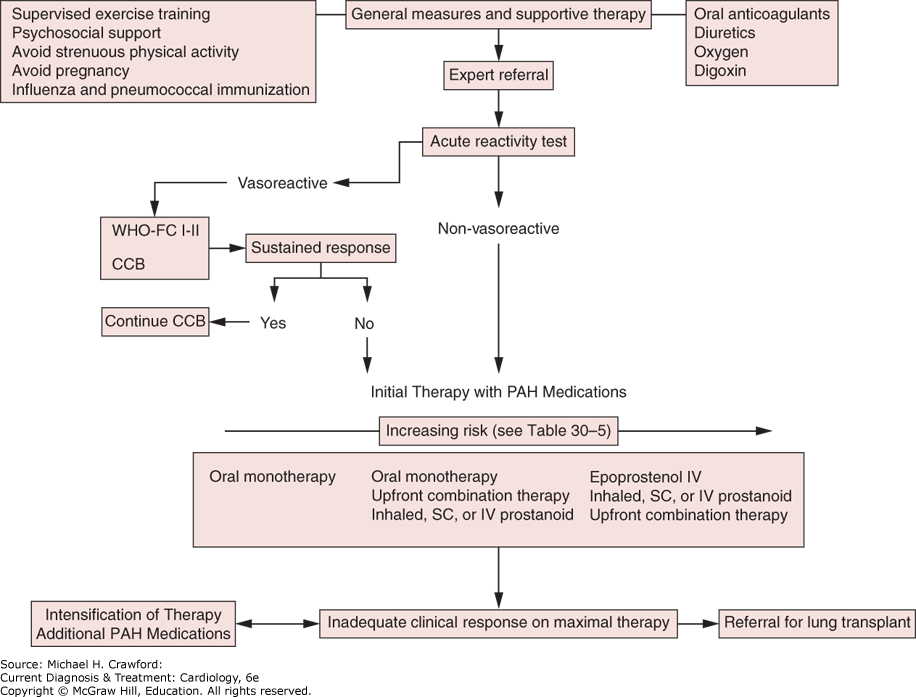
Recent improvements in medical therapy have minimized the role of invasive PAH treatments; however, lung transplantation, combined heart–lung transplantation, and atrial septostomy remain useful options in patients unresponsive to medical treatment. Mortality among PAH patients awaiting lung transplantation is high, so patients who are unresponsive to medical therapy should be referred early for lung transplant evaluation. Combined heart and lung transplantation is most often performed in patients with congenital heart disease. Atrial septostomy is performed at some expert centers as a palliative treatment for severely symptomatic PAH. The resulting right–left shunt improves left ventricular filling and CO; however, it can also worsen hypoxemia and increase the risk of paradoxical embolization.
B. Pulmonary Hypertension with Left-Sided Heart Disease
PH occurs in 25–30% of patients with LHD and is the most common cause of PH. When PH develops as a complication of LHD, it is associated with worse outcomes. In addition to optimal guideline-recommended therapy, diuretics should be used to achieve euvolemia in patients with HFrEF and HFpEF, which may require guidance with invasive assessment of PAP. Valvular heart disease and coronary artery disease should be aggressively treated in accordance with guideline recommendations.
There is currently no role for PAH therapies to patients with PH-LHD. IV prostacyclin increases mortality in patients with HFrEF and endothelin receptor antagonists worsen heart failure and increase mortality. Although short-term hemodynamic improvements are described, treatment with PDE-5 inhibitors does not yield improvement in clinically meaningful end points in HFrEF and HFpEF patients. In highly selected patients, long-term infusion of inotropes and vasodilators, ventricular assist devices, or PDE-5 inhibitors may be used to lower PAP to facilitate heart transplantation.
C. Pulmonary Hypertension Associated with Lung Disease or Hypoxemia
Lung disease is the second most common cause of PH and, in most cases, results primarily from hypoxic pulmonary vasoconstriction. Correction of hypoxemia and optimal treatment of the underlying lung disease are generally the appropriate treatments. Comorbid conditions that can cause or worsen PH from lung disease, such as HFpEF and CTEPH, are common and should be identified and treated. Inhaled treprostinil was approved to treat patients with PH from ILD in 2022. However, other PAH therapies should generally not be used because they have not been shown to improve PH from lung disease and may worsen outcomes. Sildenafil is known to worsen gas exchange, and macitentan increases exacerbations of ILD. Because development of PH in patients with lung disease is associated with increased mortality, patients who might be lung transplant candidates should undergo expedited evaluation.
D. Pulmonary Hypertension Due to Chronic Thrombotic or Embolic Disease
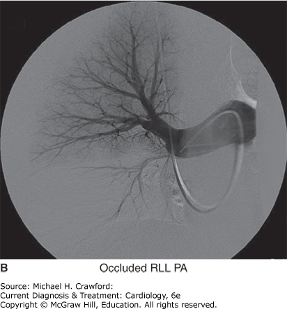
Mortality in patients with CTEPH who do not undergo surgical PEA is high, so CTEPH should be excluded in all patients undergoing evaluation for PH. Patients with suspected CTEPH should be referred to a CTEPH expert center for confirmatory conventional pulmonary angiography and surgical evaluation (Figure 30–16). At experienced centers, perioperative mortality following PEA is as low as 4.4%, and surgery may be possible in patients previously considered inoperable. Following PEA, most patients experience marked improvement in PAP, right heart function, NYHA class, and exercise capacity. Riociguat and macitentan have been approved for the treatment of inoperable CTEPH; however, it is important that this drug not be considered an alternative to surgical PEA. Percutaneous treatment of CTEPH with BPA is now offered in some CTEPH centers.
Figure 30-16. Conventional pulmonary angiography. A: Normal pulmonary angiogram. B: Pulmonary angiogram from a patient with chronic thromboembolic pulmonary hypertension demonstrating occlusion of the right lower lobe pulmonary artery (RLL PA). (Reprinted, with permission, from Barnett C, et al. Cardiac catheterization in the patient with pulmonary hypertension. In: Yuan JXJ, et al, eds. Textbook of Pulmonary Vascular Disease. New York: Springer US; 2011:13871402. Copyright © Springer Science+Business Media, 2011.)
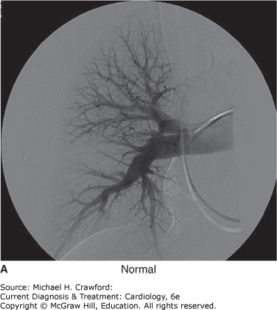
Figure 30-16.
BarnettCF, O’BrienC, MarcoTD. Critical care management of the patient with pulmonary hypertension. Eur Heart J Acute Cardiovasc Care. Jan 12 2022;11(1):7783. 34966914
GalièN, ChannickRN, FrantzRP,et al. Risk stratification and medical therapy of pulmonary arterial hypertension. Eur Respir J. Jan 2019;53(1):1801889. 30545971
GhofraniHA, SimonneauG, D’ArminiAM,et al. Macitentan for the treatment of inoperable chronic thromboembolic pulmonary hypertension (MERIT-1): results from the multicentre, phase 2, randomised, double-blind, placebo-controlled study. Lancet Respir Med. Oct 2017;5(10):785794. 28919201
HumbertM, McLaughlinV, GibbsJSR,et al. Sotatercept for the treatment of pulmonary arterial hypertension. N Engl J Med. Apr 1 2021;384(13):12041215. 33789009
RuoppNF, CockrillBA. Diagnosis and treatment of pulmonary arterial hypertension: a review. JAMA. 2022 Apr 12;327(14):13791391. 35412560
WaxmanA, JaramilloRR, ThenappanT,et al. Inhaled treprostinil in pulmonary hypertension Due to Interstitial Lung Disease. N Engl J Med. Jan 28 2021;384(4):325334. 33440084
Prognosis ⬆
PH is a progressive disorder associated with a high mortality rate. The underlying cause of the PH is a key factor in determining the prognosis and natural history of the disease (see Table 30–1). Patients with PAH associated with connective tissue disease and those with PAH and HIV have the worst prognosis. Patients with congenital heart disease, by contrast, have the best prognosis. Even for patients with PH due to lung disease or hypoxemia, survival rates vary widely. Patients with PH and emphysema have a 6-month survival of 81%, whereas patients with PH and ILD have a 6-month survival of 38%.
Clinical features and hemodynamic parameters also strongly influence survival. Markers of poor prognosis in PH include advanced functional class (NYHA III–IV) (see Table 30–2), poor exercise capacity (reduced 6MW distance), and resting hemodynamics consistent with right ventricular failure (high right atrial pressure, low CO), elevated B-type natriuretic peptide, and presence of a pericardial effusion or significant right ventricular dysfunction on echocardiogram (see Table 30–5).
BenzaRL, MaitlandMG, ElliottCG,et al. Predicting survival in patients with pulmonary arterial hypertension: The REVEAL Risk Score Calculator 2.0 and Comparison With ESC/ERS-Based Risk Assessment Strategies. Chest. Aug 2019;156(2):323337. 30772387
HumbertM, FaberHW, GhofraniHA,et al. Risk assessment in pulmonary arterial hypertension and chronic thromboembolic pulmonary hypertension. Eur Respir J. Jun 2019;53(6):1802004. 30923187