Sleep Stages & Architecture
Normal sleep involves two states: REM sleep and non-REM sleep. REM sleep is often associated with dreaming. Non-REM sleep is a period of decreased physiologic and psychological activity and is further divided into stages 1, 2, 3, and 4 on the basis of visually scored EEG patterns.
Sleep normally begins with non-REM stage 1, before progressing successively into non-REM stages 2 through 4, during which the EEG generally declines in frequency and increases in amplitude. Stages 3 and 4 of sleep, also called slow-wave sleep (SWS), are typically most intense early in the sleep period. The amount of SWS declines across the night. REM sleep is characterized by high-frequency, low-amplitude EEG; loss of muscle tone in the major antigravity muscles; and REMs (Figure 36–1).
Figure 36–1. Hypnogram of sleep stages in young versus old. (Adapted with permission from Ancoli-Israel S. All I Want Is a Good Night’s Sleep. Philadelphia: Mosby Books; 1999.)
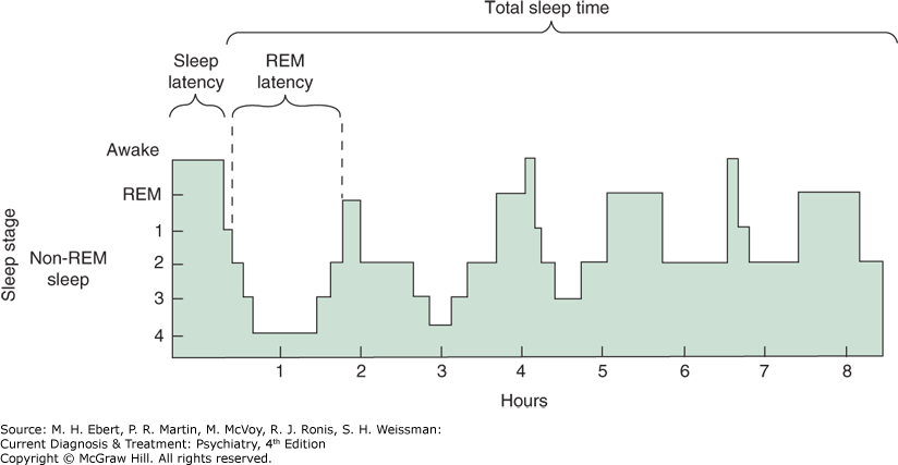 A hypnogram shows the progression through the sleep stages over 8 hours, including sleep and R E M latency.
The Neurobiology of Sleep
The neurophysiologic underpinnings of sleep and wakefulness are incompletely understood. Aspects of REM sleep such as periodic REMs and atonia are generated within the brainstem. Non-REM sleep is partially controlled by rostral brain regions such as the hypothalamus, basal forebrain, and thalamus.
A variety of neurotransmitter systems and brain regions appear to regulate sleep and wakefulness. The arousal network involves the activity of neurons containing acetylcholine, norepinephrine, serotonin, orexin (hypocretin), and dopamine (DA), whereas gamma-aminobutyric acid (GABA)-ergic mechanisms figure prominently in initiating non-REM sleep.
Sleep & Circadian Rhythms
The rhythm of sleep and wakefulness is governed by one or more internal biological “clocks,” by environmental stimuli, and by a host of processes that promote or inhibit arousal (Table 36–3). In the absence of zeitgebers (time cues such as social activities, meals, and bright lights), humans tend to self-select a sleep–wake cycle of about 25 hours from wake time to wake time. In other words, if a person lives in an experimental environment free of time cues and is allowed to go to bed and arise at will, that person will tend to go to sleep about an hour later each “night” and wake up about an hour later each “morning.” For this reason, shifts in the sleep–wake cycle activity are usually easier when the cycle is lengthened rather than shortened—in traveling west rather than east, for example—or when rotating from an afternoon to an evening work shift, rather than from an afternoon to a morning work shift.
Table 36–3 Glossary: Terms Commonly Used in the Study of Circadian Rhythms
Term | Definition |
|---|---|
Chronobiology | The study of circadian rhythms |
Circadian rhythms | Refers to biological rhythms having a cycle length of about 24 hour. Derived from Latin: circa dies, “about 1 day.” Examples include the sleep–wake cycle in humans and temperature, cortisol, and psychological variation over the 24-hour day. Characterized by exact cycle length, amplitude, and phase position. |
Phase position | Temporal relationship between rhythms or between one rhythm and the environment. For example, the maximum daily temperature peak usually occurs in the late afternoon. |
Phase-advanced rhythm | Patient retires and arises early. |
Phase-delayed rhythm | Patient retires and arises late. |
Zeitgebers | Time cues such as social activities, meals, and bright lights. |
Adapted with permission from Salzman C. Clinical Geriatric Psychopharmacology, 4th ed. Baltimore: Lippincott Williams & Wilkins; 2005.
Normally, the circadian oscillator is entrained to the 24-hour environment by zeitgebers such as social activities and meals, and especially by environmental light. Information about light reaching the retina is conveyed to the suprachiasmatic nuclei (SCN) in the anterior hypothalamus. The SCN are important oscillators that maintain the circadian rhythm of sleep–wakefulness.
In addition to synchronizing the circadian oscillator with the environment, the timing of light exposure can also shift the phase position of the oscillator (i.e., the temporal relationship between rhythms or between one rhythm and the environment). Bright light (1500 lux) in the evening hours (6–9 PM) coupled with darkness from 9 PM to 9 AM tends to cause a phase delay in sleep–wake and other biological oscillators (i.e., one would go to bed later and wake up later). In contrast, exposure to bright light in the early morning hours (5–7 AM) coupled with darkness in the evening tends to advance the phase position of the oscillator (i.e., one would go to bed earlier and wake up earlier). Furthermore, bright light during daylight hours can enhance the amplitude of the circadian rhythm, thereby demarcating the periods of both nocturnal sleep and daytime wakefulness. Bright light has been reported to have antidepressant effects in seasonal depressions occurring in the winter and in some patients with major depressive disorder or premenstrual depression.
Sleep Changes with Development & Aging
Sleep–wake states change dramatically across the life span, not only with regard to the amount of sleep but also to circadian timing. With advancing age, REM latency tends to decrease and the length of the first REM period tends to increase.
The amount of time spent each night in SWS is high in childhood, peaks in early adolescence, and gradually declines with age until it nearly disappears around the sixth decade of life. Young adults typically spend about 15–20% of total sleep time (TST) in SWS. Sleep tends to be shallower, more fragmented, and shorter in duration in middle-aged and elderly adults compared to young adults. In addition, daytime sleepiness increases. The relative amount of “shallower” stages 1 and 2 sleep tends to increase as the “deeper” stages 3 and 4`````` sleep tend to decrease. Men tend to lose SWS at an earlier age than women do.
After the age of 65, about one in three women and one in five men report that they take over 30 minutes to fall asleep. Wakefulness after sleep onset (WASO) and number of arousals increase with age, an increase that may be due at least in part to the greater incidence of sleep-related breathing disorders, PLMs, and other physical conditions in these age groups. WASO may also increase with age because older people are more easily roused by internal and external stimuli.
Changes in the circadian rhythm may lead to daytime fatigue, napping, and poor nocturnal sleep. Related to a phase-advanced temperature rhythm, elders tend to retire and arise earlier than younger adults. Psychosocial alterations can disrupt zeitgebers and light exposure. Napping also increases with age, but the TST per 24 hours does not change with age.