Usually asymptomatic. Late stages have visual field or acuity loss. History of hyphema or trauma to the glaucomatous eye can often be elicited. Glaucoma due to angle recession (not from the inciting trauma) usually takes many years to develop. Typically unilateral.
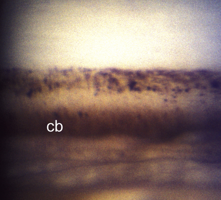
(See Figure 9.6.1.)
Critical
Glaucoma (see 9.1, PRIMARY OPEN-ANGLE GLAUCOMA) in an eye with characteristic gonioscopic findings: an uneven iris insertion, an area of torn or absent iris processes, and a posteriorly recessed iris, revealing a widened ciliary band (may be focal or extend for 360 degrees). Comparison with the contralateral eye can help identify recessed areas.
Other
The scleral spur may appear abnormally white on gonioscopy because of the recessed angle; other signs of previous trauma may be present (e.g., cataract, iris sphincter tears).
9-6.1 Angle recession glaucoma with increased width of the ciliary body band.

See 9.1, PRIMARY OPEN-ANGLE GLAUCOMA. However, miotics (e.g., pilocarpine) may be ineffective or even cause increased IOP by reduction of uveoscleral outflow. ALT and SLT are rarely effective in this condition. Surgical therapy may be necessary if IOP is uncontrolled with medications.
Both eyes are monitored closely because of high incidence of delayed open-angle glaucoma in the contralateral eye. Patients with angle recession without glaucoma are examined yearly. Those with glaucoma are examined as detailed in 9.1, PRIMARY OPEN-ANGLE GLAUCOMA.
