section name header

Symptoms

Decreased visual acuity, central/paracentral scotomata, dyschromatopsia, metamorphopsia. Typically bilateral.

Signs

(See Figure 11.35.1.)

Critical

Acute findings include a yellow-white spot in the fovea with or without surrounding granular gray pigmentation. Classic late finding is a red, sharply demarcated lesion in the fovea.

Other

Visual acuity usually ranges from 20/25 to 20/100. Amsler grid testing may reveal central or paracentral scotoma. Resolution of acute findings within several weeks may leave a variable appearance to the fovea (e.g., pigmentary disturbance, lamellar hole, normal appearance, etc). Eyes with better initial visual acuities are more likely to have unremarkable fundoscopic examinations at follow up.
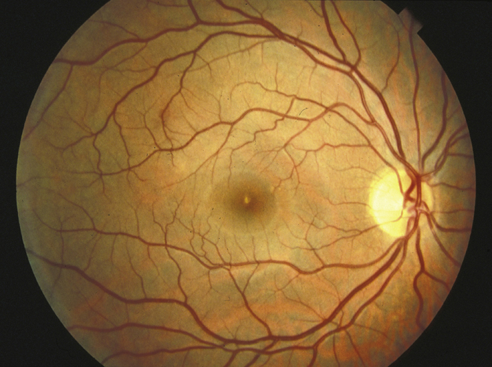
11-35.1 Solar retinopathy.

Gervasio-ch011-image059

Differential Diagnosis

Etiology

Unprotected solar eclipse viewing, sungazing (e.g., related to religious rituals, psychiatric illnesses, hallucinogenic drugs), sunbathing, vocational exposure (e.g., aviation, military service, astronomy, arc welding)

Work Up

Workup

11-35.2 Optical coherence tomography of solar retinopathy.

Gervasio-ch011-image060

Treatment

  1. Observation. Eyes with better visual acuities on initial examination tend to recover more vision. Long-term significant reduction in visual acuity is rare. However, central or paracentral scotomata may persist despite improvement in visual acuity.