section name header

Signs

Acute Phase

Critical

Marked corneal stromal blood vessels and edema.

Other

Anterior chamber cells and flare, fine keratic precipitates on the corneal endothelium, conjunctival injection.

Chronic Phase

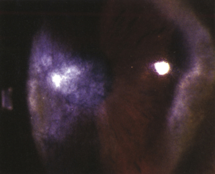
(See Figure 4.17.1.)

Deep corneal haze or scarring, corneal stromal blood vessels containing minimal or no blood (ghost vessels), stromal thinning.

4-17.1 Interstitial keratitis.

Gervasio-ch004-image013

Etiology

Most Common

HSV is the most common cause of IK (see 4.15, HERPES SIMPLEX VIRUS). Other common causes include: VZV, congenital syphilis (bilateral in 80% of cases, often occurs in the first or second decade of life, rare in first years of life. Affects both eyes within 1 year of each other, more commonly occurs as late inactive/immune-mediated disease and less often as acute/infectious disease), acquired syphilis, and tuberculosis (TB, unilateral and often sectoral).

Less Common

Epstein-Barr virus (EBV) Lyme disease, leprosy, and Cogan syndrome (autoimmune disorder characterized by bilateral IK, vertigo, tinnitus, hearing loss, and negative syphilis serologies; also associated with systemic vasculitis [e.g., polyarteritis nodosa] and typically occurs in young adults).

Work Up

Workup

For active IK or old, previously untreated IK:

  1. History: Venereal disease in the mother during pregnancy or in the patient? Hearing loss or tinnitus? Prior HSV or shingles infections?
  2. External examination: Look for saddle-nose deformity, Hutchinson teeth, frontal bossing, or other signs of congenital syphilis. Look for hypopigmented or anesthetic skin lesions and thickened skin folds, loss of the temporal eyebrow, and loss of eyelashes, as in leprosy.
  3. Slit lamp examination: Examine corneal nerves for segmental thickening, like beads on a string (leprosy). Examine the iris for nodules (leprosy) and hyperemia with fleshy, pink nodules (syphilis). Check IOP.
  4. Dilated fundus examination: Look for classic salt-and-pepper chorioretinitis or optic atrophy of syphilis.
  5. Laboratory evaluation: Fluorescent treponemal antibody absorption (FTA-ABS) or treponemal-specific assay (e.g., microhemagglutination-Treponema pallidum [MHA-TP]) for prior exposure; Venereal Disease Research Laboratory test (VDRL) or rapid plasma reagin (RPR) for disease activity. See 12.12, SYPHILIS.
  6. Purified protein derivative (PPD) or interferon-gamma release assay (IGRA) (e.g., QuantiFERON-TB Gold),
  7. Lyme serology if in endemic region.
  8. Chest radiograph or chest CT if negative FTA-ABS (or MHA-TP) or positive PPD or IGRA.
  9. Consider erythrocyte sedimentation rate, rheumatoid factor, complete blood count, and EBV antibody.

Treatment

  1. Acute disease:
    • Topical cycloplegic drop (e.g., cyclopentolate 1% t.i.d. or atropine 1% b.i.d.).
    • Topical steroid (e.g., prednisolone acetate 1% q2–6h depending on the degree of inflammation).
    • Treat any underlying disease.
  2. Old inactive disease with central scarring:
    • RGP or scleral contact lens if vision decreased from irregular astigmatism
    • Corneal transplantation may improve vision if minimal amblyopia is present.
  3. Recently inactive or old inactive disease:
    • If the treponemal-specific assay or FTA-ABS is positive and the patient has active or untreated syphilis, or if the VDRL or RPR titer is positive and has not declined the expected amount after treatment, then treatment for syphilis is indicated. See 12.12, SYPHILIS.
    • If PPD or IGRA is positive and the patient is <35 years and has not been treated for TB in the past, or there is evidence of active systemic TB (e.g., positive finding on chest radiograph), then refer the patient to an internist and infectious disease specialist for TB treatment.
    • If Cogan syndrome is present, refer the patient to an otolaryngologist and rheumatologist.
    • If Lyme antibody and titers are positive, treat per 13.4, LYME DISEASE.

Follow Up

  1. Acute disease: Every 3 to 7 days initially, and then every 2 to 4 weeks. The frequency of steroid administration is slowly reduced as the inflammation subsides over the course of months (may take years). IOP is monitored closely and reduced with medications based on the degree of IOP elevation and overall health of the optic nerve (see 9.7, INFLAMMATORY OPEN ANGLE GLAUCOMA).
  2. Old inactive disease: Yearly follow up, unless treatment is required for underlying etiology.

IK is broadly defined as any non-ulcerating inflammation of the corneal stroma without epithelial or endothelial involvement, but often with neovascularization. IK is the common end-point of many corneal diseases.