section name header

Signs

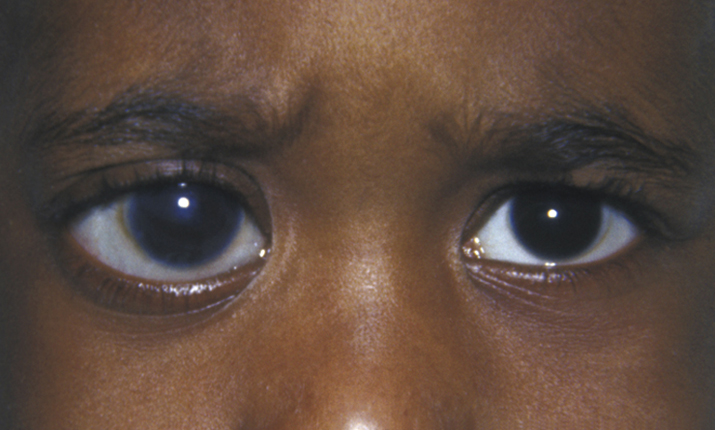
(See Figure 8.11.1.)

Critical

Enlarged globe and corneal diameter (horizontal corneal diameter >12 mm before 1 year of age is suggestive), corneal edema, Haab striae (curvilinear tears in Descemet membrane of the cornea, with scalloped edges with or without associated stromal haze), increased cup/disc ratio, high intraocular pressure (IOP), axial myopia, commonly bilateral (80%). Classic findings are tearing, photophobia, blepharospasm, corneal clouding, and a large eye (buphthalmos).

Other

Corneal stromal scarring or opacification; high iris insertion on gonioscopy; other signs of iris dysgenesis, including heterochromia, may exist.

8-11.1 Buphthalmos of right eye in congenital glaucoma.
Gervasio-ch008-image012

Differential Diagnosis

  • Megalocornea: Bilateral horizontal corneal diameter usually >13 mm, with normal corneal thickness and endothelium, IOP, and cup/disc ratio. Radial iris transillumination defects may be seen. Usually X-linked recessive (boys affected, female carriers may have greater than normal corneal diameters) and may be associated with developmental delay (Neuhauser syndrome, autosomal recessive).
  • Trauma from forceps during delivery: May produce tears in the Descemet membrane and localized corneal edema; tears are typically vertical or oblique. Corneal diameter is normal. Usually unilateral and must have history of forceps use to make diagnosis.
  • Congenital hereditary endothelial dystrophy: Bilateral full-thickness corneal edema at birth with a normal corneal diameter and axial length. IOP may be falsely elevated by increased corneal thickness and hysteresis but true associated infantile glaucoma has been reported. See 4.25, CORNEAL DYSTROPHIES.
  • Posterior polymorphous dystrophy: Can present in infancy as bilateral but asymmetric cloudy edematous corneas with characteristic endothelial abnormalities. Normal corneal diameter, axial length, and IOP, but carries lifetime glaucoma risk. Abnormal endothelium may be seen in one parent. See 4.25, CORNEAL DYSTROPHIES.
  • Mucopolysaccharidoses and cystinosis: Some inborn errors of metabolism produce cloudy corneas in infancy or early childhood, usually not at birth. The corneal diameter and axial length are normal. IOP is rarely elevated and, if so, usually later in childhood. Always bilateral.
  • Nasolacrimal duct obstruction: No photophobia, clear cornea, normal corneal size and axial length, normal IOP. See 8.10, CONGENITAL NASOLACRIMAL DUCT OBSTRUCTION.
  • Large eye without other signs of glaucoma can be seen in overgrowth syndromes (e.g., hemihypertrophy) and phakomatoses (e.g., neurofibromatosis, Sturge–Weber) in the absence of glaucoma (although these diagnoses may carry a high risk of glaucoma). May also be autosomal dominant variant without glaucoma.

Etiology

Common

  • Primary congenital glaucoma: Not associated with other ocular or systemic disorders. Diagnosed after other causes of glaucoma have been ruled out. Caused by incomplete differentiation of the trabecular meshwork during embryogenesis (e.g., goniodysgenesis).
  • Glaucoma following cataract surgery: Most common form of pediatric glaucoma. Typically in older children. All children undergoing cataract surgery are at lifelong risk.

Less Common

  • Sturge–Weber syndrome: Usually unilateral (90%); ipsilateral port-wine mark almost always involving eyelid(s), cerebral calcifications/atrophy, and seizures/developmental delay (central nervous system may not be involved at all); not familial. See 13.13, PHAKOMATOSES.

Rare

  • Other anterior segment dysgeneses: Axenfeld–Rieger spectrum, Peters anomaly, others. See 8.12, DEVELOPMENTAL ANTERIOR SEGMENT AND LENS ANOMALIES/DYSGENESIS.
  • Lowe syndrome (oculocerebrorenal syndrome): Cataract, glaucoma, developmental delay, and renal disease; X-linked recessive.
  • Congenital rubella: Glaucoma, cataract, “salt-and-pepper” retinopathy, hearing and cardiac defects (usually peripheral pulmonic stenosis).
  • Aniridia: Absence of most of iris, often with only a rudimentary iris stump visible on gonioscopy. Associated with cataracts, glaucoma, macular hypoplasia, and nystagmus. See 8.12, DEVELOPMENTAL ANTERIOR SEGMENT AND LENS ANOMALIES/DYSGENESIS.
  • Others: Neurofibromatosis, PFV, Weill–Marchesani syndrome, Rubinstein–Taybi syndrome, covert trauma, steroid-induced infantile glaucoma, complication of ROP, and intraocular tumors.

Work Up

Workup
  1. History: Other systemic abnormalities? Rubella infection during pregnancy? Birth trauma? Family history of congenital glaucoma?
  2. Ocular examination, including a visual acuity assessment of each eye separately, measurement of horizontal corneal diameters (measured with calipers or templates), IOP measurement, and a slit lamp or portable slit lamp examination to evaluate for corneal edema and Haab striae. Retinoscopy to estimate refractive error looking for axial myopia. A dilated fundus examination is performed to evaluate the optic disc and retina if able to view through cornea.
  3. EUA is performed in cases too difficult to evaluate in the office and in those for whom surgical treatment is considered. Horizontal corneal diameter, IOP measurement, pachymetry, retinoscopy, gonioscopy, and ophthalmoscopy are performed. Axial length is measured with ultrasound (A-scan). At 40 gestational weeks, normal mean axial length is 17 mm. This increases to 20 mm on average by age 1 year. Axial length progression may also be monitored by successive cycloplegic refractions or serial ultrasounds. Disc photos may be taken.
NOTE:

IOP may be reduced by general anesthesia, particularly halothane (sevoflurane or desflurane less likely), and over ventilation (low end-tidal CO2); IOP may be elevated with ketamine hydrochloride, succinylcholine, endotracheal intubation (for 2 to 5 minutes), pressure from the anesthetic mask, speculum use, or inadequate ventilation with elevated end-tidal CO2.

Treatment

Definitive treatment is surgical, particularly in primary congenital glaucoma. Medical therapy is utilized as a temporizing measure before surgery and to help clear the cornea in preparation for possible goniotomy.

  1. Medical:
    • Oral carbonic anhydrase inhibitor (e.g., acetazolamide, 15 to 30 mg/kg/d in three or four divided doses): Most effective.
    • Topical carbonic anhydrase inhibitor (e.g., dorzolamide or brinzolamide b.i.d.): Less effective; better tolerated.
    • Topical beta-blocker (e.g., levobunolol or timolol, 0.25% if <1 year old or 0.5% if older b.i.d.): Important to avoid in asthma patients (betaxolol preferable).
    • Prostaglandin analogs (e.g., latanoprost q.h.s.).
  2. Surgical: Nasal goniotomy (incising the trabecular meshwork with a blade or needle under gonioscopic visualization) is the procedure of choice, although some surgeons initially recommend trabeculotomy. Miotics are sometimes used to constrict the pupil before a surgical goniotomy. If the cornea is not clear, trabeculotomy (opening the Schlemm canal from a scleral approach ab externo into the anterior chamber) or endoscopic goniotomy can be performed. If the initial goniotomy is unsuccessful, a temporal goniotomy may be tried. Trabeculectomy or tube shunt may be performed following failed angle incision operations. Cyclodestruction of the ciliary processes through cyclophotocoagulation or cryotherapy may also be an option to decrease aqueous production in certain circumstances.
NOTE:

Brimonidine is contraindicated in children under the age of 1 year because of the risk of apnea/hypotension/bradycardia/hypothermia from blood–brain permeability. Caution should be used in children under 5 years old or <20 kg or intracranial pathology (such as Sturge–Weber syndrome).

NOTE:

Amblyopia is the most common cause of visual loss in pediatric glaucoma and should be treated appropriately. See 8.7, AMBLYOPIA.

Follow Up

  1. Repeated examinations, under anesthesia as needed, to monitor corneal diameter and clarity, IOP, cup/disc ratio, and refraction/axial length.
  2. These patients must be followed throughout life to monitor for progression.
  3. Other forms of pediatric glaucoma in older children include uveitic glaucoma, traumatic glaucoma, juvenile open-angle glaucoma (autosomal dominant), and others.