section name header

Symptoms

Often asymptomatic early, then decreased vision, abnormal color vision, reduced dark adaptation.

Signs

Critical

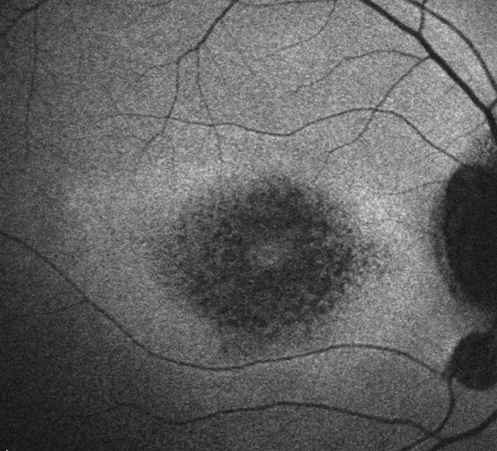
Bull’s eye macula (ring of depigmentation surrounded by a ring of increased pigmentation), loss of foveal reflex.

Other

Increased pigmentation in the macula, arteriolar narrowing, vascular sheathing, peripheral pigmentation, decreased color vision, CME, visual field abnormalities (central, paracentral, or peripheral scotoma), abnormal ERG and EOG, and abnormal dark adaptation. Whorl-like corneal changes may also be observed.

Major Risk Factors

  • Chloroquine daily dosage: >2.3 mg/kg real weight
  • Hydroxychloroquine daily dosage: >5.0 mg/kg real weight
  • Duration of use: >5 years, assuming no other risk factors
  • Renal disease, tamoxifen use, comorbid macular disease

Differential Diagnosis

Differential Diagnosis of Bull’s Eye Maculopathy

Reference(s)

Marmor MF, Kellner U, Lai TY, et al; American Academy of Ophthalmology. Recommendations on screening for chloroquine and hydroxychloroquine retinopathy (2016 Revision). Ophthalmology. 2016;123(6):1386-1394.

Treatment

Discontinue the medication in conjunction with the prescribing physician if signs of toxicity develop.

Baseline Workup

Baseline evaluation should be performed within the first year of starting the medication.

  1. Best corrected visual acuity.
  2. Ophthalmoscopic examination, including dilated fundus examination with particular attention to any pigmentary alterations.
  3. Consider posterior pole fundus photographs.
  4. Consider visual fields and OCT if maculopathy is present.

Follow Up

After 5 years of medication use (sooner in presence of major risk factors), begin annual screening:

  1. Automated visual fields: Preferably white SITA testing and 10-2 pattern for non-Asians. 24-2 or 30-2 pattern recommended for Asian patients in whom toxicity often manifests in the more peripheral macula.
  2. Spectral domain OCT: Parafoveal photoreceptor layer thinning and/or disruption of outer retinal layers (“flying saucer sign”), RPE atrophy, loss of foveal contour. Consider wide angle scans including vascular arcades in Asian patients.
  3. Additional tools that may be used as available or in suspect cases include multifocal ERG and FAF (see Figure 11.32.1)

11-32.1 Fundus autofluorescence of hydroxychloroquine toxicity.

Gervasio-ch011-image057
NOTE:

Once ocular toxicity develops, it usually does not regress even if the drug is withdrawn. In fact, new toxic effects may develop, and old ones may progress even after the chloroquine/hydroxychloroquine has been discontinued.