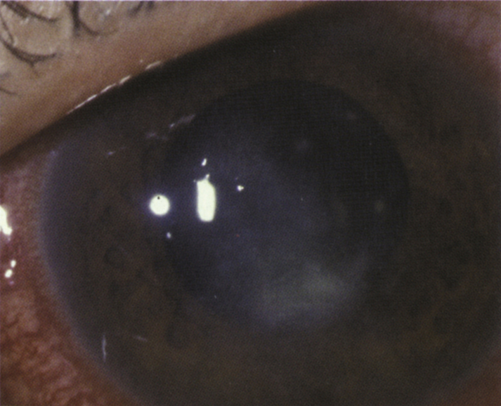
Pain, photophobia, redness, tearing, discharge, foreign body sensation. Often history of minor trauma particularly with vegetable matter (e.g., a tree branch), contact lens wear, chronic eye disease, and/or a history of poor response to conventional antibacterial therapy. Usually more indolent than bacterial keratitis.
WorkupSee 4.11, BACTERIAL KERATITIS for complete workup and culture procedure.
- Whenever smears and cultures are done, include a Gram, Giemsa, calcofluor white, or KOH stain for organisms; periodic acid-Schiff, Gomori methenamine silver, and hematoxylin and eosin (H&E) stains can also be used. Scrape deep into the edge of the ulcer for material. See Appendix 8, CORNEAL CULTURE PROCEDURE.
- If an infectious etiology is still suspected despite negative cultures, consider a corneal biopsy to obtain further diagnostic information.
- Consider cultures and smears of contact lens case and solution.
- Sometimes all tests are negative, yet the disease continues to progress and therapeutic penetrating keratoplasty (PK) is necessary for diagnosis and treatment.
Corneal infiltrates and ulcers of unknown etiology are treated as bacterial until proven otherwise (see 4.11, BACTERIAL KERATITIS). If the stains or cultures indicate a fungal keratitis, institute the following measures:
- Natamycin 5% drops (especially for filamentous fungi), amphotericin B 0.15% drops (especially for Candida), and/or topical fortified voriconazole 1% initially q1–2h around the clock, then taper over the next several weeks based on response to therapy, (see Appendix 9, FORTIFIED TOPICAL ANTIBIOTICS/ANTIFUNGALS).
- Cycloplegic drop (e.g., cyclopentolate 1% t.i.d.; atropine 1% b.i.d. to t.i.d. is recommended if hypopyon, fibrin or significant anterior chamber reaction is present).
- No topical steroids. If the patient is currently taking steroids, they should be tapered rapidly and discontinued.
- Consider adding oral antifungal agents (e.g., fluconazole or itraconazole 200 to 400 mg p.o. loading dose, then 100 to 200 mg p.o. daily, posaconazole 300 mg p.o. b.i.d. for 1 day then 300 mg p.o. q.d., or voriconazole 200 mg p.o. b.i.d.). Oral antifungal agents are often used for deep corneal ulcers or suspected fungal endophthalmitis.
- Consider epithelial debridement to facilitate the penetration of antifungal medications. Topical antifungals do not penetrate the cornea well, especially through an intact epithelium. When culture results and sensitivities are known, intrastromal depot injections of amphotericin (10 mcg/0.1 mL) or voriconazole (voriconazole 50 mcg/0.1 mL) can also be considered.
- Measure IOP (preferably with Tono-Pen). Treat elevated IOP if present (see 9.7, INFLAMMATORY OPEN ANGLE GLAUCOMA).
- Eye shield, without patch, in the presence of corneal thinning.
- Admission to the hospital may be necessary if patient compliance is an issue. It may take weeks to achieve complete healing.
- If unable to adequately control the infection despite prolonged treatment, a therapeutic penetrating keratoplasty can be considered, ideally before the infection reaches the limbus. Intracameral antifungal medications (e.g., voriconazole 50 mcg/0.1 mL or amphotericin 10 mcg in 0.1 mL) at the time of therapeutic keratoplasty should be considered. Anterior lamellar keratoplasty is relatively contraindicated because there is a high risk of recurrence of infection.
 NOTE: NOTE: |
Natamycin is the only commercially available topical antifungal agent; all others must be compounded. |
Patients are reexamined daily at first. However, the initial clinical response to treatment in fungal keratitis is much slower compared to bacterial keratitis. Stability of infection after initiation of treatment is often a favorable sign. Unlike bacterial ulcers, epithelial healing in fungal keratitis is not always a sign of positive response. Fungal infections in deep corneal stroma may be recalcitrant to therapy. These ulcers may require weeks to months of treatment, and therapeutic corneal transplantation may be necessary for infections that progress despite maximal medical therapy or corneal perforation.
