section name header

Symptoms

Glare and blurred vision, worse on awakening. May progress to severe pain due to ruptured bullae. Symptoms usually develop in the fifth and sixth decades.

Signs

Critical

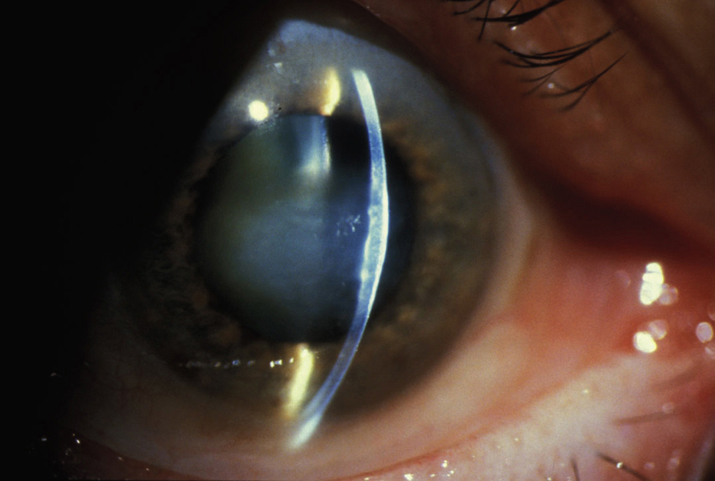
Cornea guttae with stromal +/- epithelial edema (see Figure 4.26.1). Bilateral, but may be asymmetric.

NOTE:

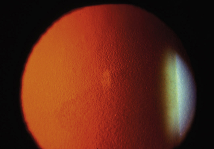
Central cornea guttae without stromal edema is called endothelial dystrophy (see Figure 4.26.2). This condition may progress to Fuchs dystrophy over years.

4-26.2 Corneal guttae in endothelial dystrophy.

Gervasio-ch004-image031

4-26.1 Corneal edema secondary to Fuchs endothelial dystrophy.

Gervasio-ch004-image030

Other

Fine pigment dusting on the endothelium, central epithelial edema and bullae, folds in Descemet membrane, stromal edema, subepithelial haze, or scarring.
Differential Diagnosis

  • Postsurgical corneal edema (also known as aphakic or pseudophakic bullous keratopathy): History of cataract surgery or other anterior segment surgery (e.g., tube shunt). See 4.27, APHAKIC BULLOUS KERATOPATHY/PSEUDOPHAKIC BULLOUS KERATOPATHY.
  • Congenital hereditary endothelial dystrophy: Bilateral corneal edema at birth. See 4.25, CORNEAL DYSTROPHIES.
  • Posterior polymorphous corneal dystrophy: Seen early in life. Corneal endothelium shows grouped vesicles, geographic gray lesions, or broad bands. Occasionally corneal edema. See 4.25, CORNEAL DYSTROPHIES.
  • Iridocorneal endothelial syndrome: “Beaten metal” corneal endothelial appearance, with corneal edema, peripheral anterior synechiae, increased IOP, variable iris thinning, corectopia, and polycoria. Typically unilateral, in young to middle-aged adults. See 9.15, IRIDOCORNEAL ENDOTHELIAL SYNDROME.

Work Up

Workup
  1. History: Previous surgery?
  2. Slit lamp examination: Cornea guttae are best seen with retroillumination. Fluorescein staining may demonstrate intact or ruptured bullae.
  3. Measure IOP.
  4. Corneal pachymetry to determine the central corneal thickness.
  5. Consider specular microscopy to evaluate the endothelial cells although may be difficult to image the endothelial layer through an edematous cornea and in between guttate changes.
  6. Consider corneal tomography to help monitor progression of disease.

Treatment

  1. Topical sodium chloride 5% drops q.i.d. and ointment q.h.s.
  2. May gently blow warm air from a hair dryer at arm’s length toward the eyes for a few minutes every morning to dehydrate the cornea.
  3. IOP reduction if indicated; also may help with corneal edema.
  4. Ruptured corneal bullae are painful and should be treated as recurrent erosions (see 4.2, RECURRENT CORNEAL EROSION).
  5. Surgery: Endothelial keratoplasty (Descemet membrane endothelial keratoplasty [DMEK] or Descemet stripping endothelial keratoplasty [DSEK]) is usually indicated when visual acuity decreases due to corneal edema; PK is indicated if significant anterior stromal scarring is present. Descemet stripping only (DSO) may be an option for milder forms of Fuchs endothelial dystrophy.

Follow Up

Every 3 to 12 months to check IOP and assess corneal edema. The condition progresses very slowly, and visual acuity typically remains good until stromal edema, epithelial edema, and/or corneal scarring develop.