Decreased visual acuity, central/paracentral scotomata, dyschromatopsia, metamorphopsia. Typically bilateral.
(See Figure 11.35.1.)
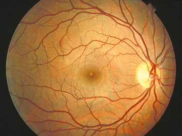
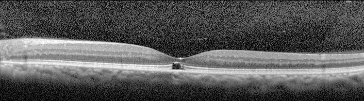
Acute findings include a yellow-white spot in the fovea with or without surrounding granular gray pigmentation. Classic late finding is a red, sharply demarcated lesion in the fovea.
Visual acuity usually ranges from 20/25 to 20/100. Amsler grid testing may reveal central or paracentral scotoma. Resolution of acute findings within several weeks may leave a variable appearance to the fovea (e.g., pigmentary disturbance, lamellar hole, normal appearance). Eyes with better initial visual acuities are more likely to have unremarkable fundoscopic examinations at follow-up.
Macular hole or vitreomacular traction: See 11.25, Vitreomacular Adhesion/Vitreomacular Traction/Macular Hole.
Idiopathic macular telangiectasia type 2: May have OCT findings similar to those seen in chronic solar retinopathy, although on IVFA juxtafoveal capillary telangiectasis is seen with leakage. May be complicated by CNV.
Intraretinal cysts, as for example in chronic CME, but this can be differentiated by OCT.
Pattern dystrophy: Adult-onset foveomacular dystrophy. See 11.31, Best Disease (Vitelliform Macular Dystrophy).
Unprotected solar eclipse viewing, sungazing (e.g., related to religious rituals, psychiatric illnesses, hallucinogenic drugs), sunbathing, vocational exposure (e.g., aviation, military service, astronomy, arc welding).
History: Eclipse viewing or sungazing? Work exposure? Photosensitizing medications?
Complete ocular examination, including a dilated fundus examination and careful inspection of the macula with a slit lamp using a handheld lens.
Amsler grid testing may identify central or paracentral scotomata.
IVFA typically shows a window defect late in the disease course.
OCT findings in the acute setting include hyporeflectivity at the level of the RPE and occasional hyperreflectivity of the injured neurosensory retina. In the chronic stage, a central hyporeflective defect at the level of the photoreceptor inner segmentouter segment junction is seen (see Figure 11.35.2).