section name header

Symptoms

Decreased vision, scotoma, metamorphopsia, or asymptomatic.

Signs

(See Figure 11.31.1.)

Figure 11.31.1: Best disease.

Rapuano9781975243722-ch011_f056.jpg

Critical

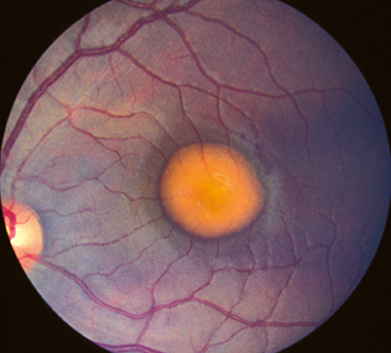
Yellow, round, subretinal lesion(s) likened to an egg yolk (lipofuscin) or in some cases to a pseudohypopyon. Typically bilateral and located in the fovea, measuring approximately one to two disc areas in size. Likely present at birth, though may not be detected until examination is performed. Ten percent of lesions  are multiple and extrafoveal. ERG is typically normal, while EOG is abnormal, showing severe loss of the light response.

Other

The lesions may degenerate, and patients may develop macular CNV (20% of patients), hemorrhage, and atrophic scarring. In the scar stage, it may be indistinguishable from AMD. Hyperopia is common due to shortened axial length along with angle-closure glaucoma; may also have esophoria or esotropia.

Inheritance

Autosomal dominant with variable penetrance and expression. Due to a mutation of the BEST1 gene. An autosomal recessive form with many small vitelliform lesions is also occasionally seen and diagnosed earlier in age than the autosomal dominant form.

Differential Diagnosis

Workup

  1. Family history. It is often helpful to examine family members.

  2. Complete ocular examination, including a dilated fundus examination, carefully inspecting the macula with a slit lamp and a handheld lens.

  3. EOG is highly specific and can be used to confirm the diagnosis or to detect the carrier state of the disease. Carriers may have normal fundi but an abnormal EOG, though EOG is used less in the era of genetic testing.

  4. Genetic testing to confirm mutation of BEST1 gene.

  5. Consider IVFA or OCTA and OCT to detect the presence of or to delineate CNV.

Treatment

There is no effective treatment for the underlying disease. Treatment for CNV is controversial because it may heal without devastating visual loss. In the era of intravitreal anti-VEGF agents, these are now typically first-line agents for CNV, but PDT and focal laser may also play a potential role. Additionally, due to development of subretinal hemorrhage with mild trauma, polycarbonate lenses are recommended at all times and especially while playing sports. See 11.17, Neovascular or Exudative (Wet) Age-Related Macular Degeneration, for detailed treatment options for CNV.

Follow-Up

Patients with CNV should be treated promptly. Otherwise, there is no urgency in seeing patients with this disease. Patients are given an Amsler grid (see Appendix 4, Amsler Grid), instructed on its use, and told to return immediately if a change is noted.