Also see 9.4, Acute Angle Closure Attack.
Perform prelaser peripheral iridotomy (LPI) gonioscopy to assess the baseline angle.
Inform the patient that they may experience ghost imaging after LPI due to the newly created iris defect. Preference for creation of the LPI at 3 and 9 o’clock to avoid the eyelid margin and prismatic tear film effect theorized to cause ghost images. Superior LPI discouraged even if completely covered by upper eyelid due to high rate of postoperative dysphotopsias.
Pretreat the eye with one drop each of apraclonidine 1% and pilocarpine (1% for lightly pigmented irides and 2% for darkly pigmented). As an alternative to pilocarpine, some ophthalmologists prefer to shine a bright light into the fellow eye immediately before engaging the laser or to employ bright ambient light. This allows for physiologic constriction of the operative pupil.
Place an Abraham yttrium–aluminum–garnet (YAG) iridotomy contact lens cushioned with 2.5% hydroxypropyl methyl cellulose or lidocaine gel, positioning the magnification button above the anticipated site of iris penetration.
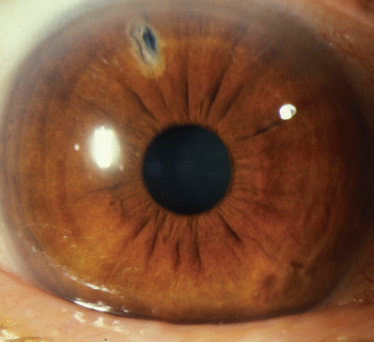
Focus the YAG beam on the predetermined iris location (see #2, above). Focus within an iris crypt if possible (see Figure A.15.1).
Engage the laser. There will be a gush of posterior iris pigment when the iris is completely penetrated. If not penetrated, advance the YAG beam to refocus on the newly created crater. Re-engage the laser until the iris is completely penetrated.
Administer one drop of prednisolone 1% and apraclonidine 1% after laser treatment.
Treat inflammation with prednisolone 1% q.i.d. for 4 to 7 days. If the LPI required a significant amount of power (e.g., more than six triple shots), taper the steroids before discontinuation to prevent rebound inflammation.
Have the patient return within 1 to 2 weeks for IOP measurement, iridotomy evaluation, and gonioscopy.