section name header

Symptoms

Decreased vision, floaters, and pain that is often bilateral. Patients typically have a history of recent hospitalization, recent abdominal surgery, being immunocompromised, possessing a long-term indwelling line or catheter (e.g., for hyperalimentation, hemodialysis, or antibiotics), or using intravenous drugs.

Signs

(See Figure 12.18.1.)

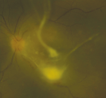
Figure 12.18.1: Candida chorioretinitis with vitreous involvement.

Rapuano9781975243722-ch012_f021.jpg

Critical

Discrete, multifocal, yellow-white, choroidal to chorioretinal fluffy lesions from one to several disc diameters in size. With time, the lesions increase in size, break into the vitreous, and appear as a “string of pearls,” especially in the case of Candida. Aspergillus has a predisposition for the subretinal space. 

Other

Vitreous cell and haze, vitreous abscesses, retinal hemorrhages with or without pale centers, AC cells, and hypopyon. A retinal detachment may develop.

Differential Diagnosis

The following should be considered in immunocompromised patients.

Workup

  1. History: History of bacteremia or fungemia? Underlying medical conditions? Medications? Indwelling catheter? Intravenous drug use? Other risk factors for immunocompromised state?

  2. Skin examination for signs of intravenous drug injection.

  3. Most clinicians recommend that all patients with fungemia have a complete dilated fundus examination (ideally within 72 hours), as ocular involvement may be asymptomatic. A repeat dilated fundus examination is recommended 2 weeks after the initial negative examination.

  4. Blood, urine, and catheter tip fungal cultures; these often need to be repeated several times and may be negative despite ocular candidiasis. Blood cultures may need to be held for a full 7 days to assess for fungal species.

  5. Consider vitrectomy to obtain a specimen and remove opacified vitreous. Cultures and smears can confirm the diagnosis. Amphotericin B 5 to 10 µg in 0.1 mL or voriconazole 50 to 100 µg in 0.1 mL is injected into the vitreous cavity after the procedure.

Treatment

  1. Suspected fungal endophthalmitis without a clear source should be considered evidence of a disseminated infection and requires further systemic evaluation and workup.

  2. Hospitalize all unreliable patients, systemically ill patients, or those with moderate to severe vitreous involvement.

  3. An infectious disease specialist should be consulted for systemic workup to evaluate for a source and other sites of involvement.

  4. Typically, chorioretinitis without vitreous involvement can be successfully treated with systemic therapy alone with one of the following regimens: Fluconazole 800 mg p.o. loading dose followed by 400 to 800 mg p.o. daily. Alternatively, voriconazole 400 mg i.v. b.i.d. daily for two doses followed by 300 mg i.v. or p.o. b.i.d. may also be considered in fluconazole-resistant species. For fluconazole- and voriconazole-resistant species, liposomal amphotericin B (3 to 5 mg/kg i.v. daily) is recommended. Therapy should be guided by cultures and sensitivities. Other agents that may be used include caspofungin, itraconazole, and micafungin.

  5. Intravitreal injection of antifungal agents as above (voriconazole or amphotericin B) if there is vitreous involvement. Depending on the response and location of retinal involvement (anterior versus posterior), injections may be repeated.

  6. Topical cycloplegic agent (e.g., atropine 1% b.i.d. to t.i.d.).

  7. See 9.7, Uveitic Glaucoma, for IOP control.

Follow-Up

  1. Patients are seen daily early on. Visual acuity, IOP, and the degree of AC and vitreous inflammation are assessed.

  2. Patients receiving azole antifungals require liver function tests every 1 to 2 weeks and as clinically indicated. Patients receiving amphotericin require monitoring of electrolytes, kidney function, and CBC as directed by an infectious disease specialist.