section name header

General Information

Definition

Symptoms

Decreased vision, double vision that persists when covering one eye (monocular diplopia).

Signs

(See Figure 13.2.1.)

Figure 13.2.1: Ectopia lentis.

Rapuano9781975243722-ch013_f002.jpg

Critical

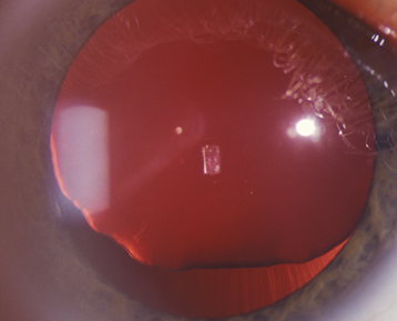
Decentered or displaced lens, iridodonesis (quivering of the iris), and phacodonesis (quivering of the lens).

Other

Change in refractive error, marked astigmatism, cataract, angle-closure glaucoma as a result of pupillary block, vitreous in the anterior chamber, and asymmetry of the anterior chamber depth.

Etiology

Workup

  1. History: Trauma? Family history of disorders listed above? Systemic illness (e.g., syphilis)? Neurologic symptoms (e.g., seizures)?

  2. Determine whether the condition is unilateral or bilateral. Determine direction of lens displacement and evaluate for subtle phacodonesis by observing the lens during back-and-forth saccadic eye movements. Check for pseudoexfoliation. Evaluate for acute or remote signs of ocular trauma including hyphema, angle recession, iridodialysis, cyclodialysis, retinal tears, and detachments.

  3. Systemic examination: Evaluate stature, extremities, hands, and fingers; often in conjunction with an internist, including blood and urine tests to rule out homocystinuria and echocardiography to rule out aortic aneurysms in patients with possible Marfan syndrome. Consider genetic testing when appropriate and available.

  4. Syphilis screening tests (rapid plasma reagin [RPR] or venereal disease research laboratory [VDRL] and fluorescent treponemal antibody absorption [FTA-ABS] or treponemal-specific assay if indicated).

Treatment

  1. Lens dislocated into the anterior chamber.

    • Dilate the pupil, place the patient on his or her back, and attempt to replace the lens into the posterior chamber by head manipulation. It may be necessary to indent the cornea after topical anesthesia with a gonioprism or cotton swab to reposition the lens. After the lens is repositioned in the posterior chamber, constrict the pupil with pilocarpine 0.5% to 1% q.i.d. and perform a peripheral laser iridotomy to prevent pupillary block.

      or

    • Surgically remove the lens and consider placing an IOL (preferred treatment if significant cataract, corneal decompensation, prior treatment failure, recurrent dislocation, or compliance issues with pilocarpine).

  2. Lens dislocated into the vitreous.

    • Lens capsule intact, patient asymptomatic, no signs of inflammation: Observation versus pars plana lensectomy and possible IOL placement.

    • Lens capsule broken with intraocular inflammation: Pars plana lensectomy with possible IOL placement.

  3. Subluxation.

    • Asymptomatic or stable refractive error: Observe in adults. Timely refractive correction to prevent amblyopia in children.

    • Uncorrectable astigmatism, unstable refractive errors, or monocular diplopia: Surgical removal of the lens and possible IOL placement.

    • Symptomatic cataract: Options include surgical removal of the lens, mydriasis (e.g., atropine 1% daily) and aphakic correction (i.e., contact lens if the other eye is phakic or pseudophakic to prevent anisometropia), pupillary constriction (e.g., pilocarpine 4% gel q.h.s.) and phakic correction.

  4. Pupillary block: Treatment is identical to that for aphakic pupillary block. See 9.15, Postoperative Glaucoma.

  5. If Marfan syndrome suspected, refer the patient to a cardiologist for an annual echocardiogram and management of any cardiac-related abnormalities. Prophylactic systemic antibiotics may be needed if the patient undergoes surgery (or a dental procedure) to prevent endocarditis.

  6. If homocystinuria is suspected: Refer to an internist. The usual therapy consists of:

    • Pyridoxine (vitamin B6) daily oral supplementation and a methionine-restricted, cysteine-supplemented diet.

    • Avoid surgery, if possible, because of the risk of thromboembolic complications. If surgical intervention is necessary, anticoagulant therapy is indicated in conjunction with internist.

Follow-Up

Depends on the etiology, degree of subluxation or dislocation, and symptoms.