Definition
A systemic disorder in which grayish-white exfoliation material, along with pigment released from the iris sphincter region, block the TM and raise the IOP. Up to 44% of patients with pseudoexfoliation syndrome may require glaucoma treatment over time. Pseudoexfoliation is the most common secondary glaucoma in those of European descent, but can be seen in nearly all ethnic groups.
Usually asymptomatic in its early stages. Unlike POAG, more often asymmetric at presentation.
(See Figures 9.10.1 and 9.10.2.)
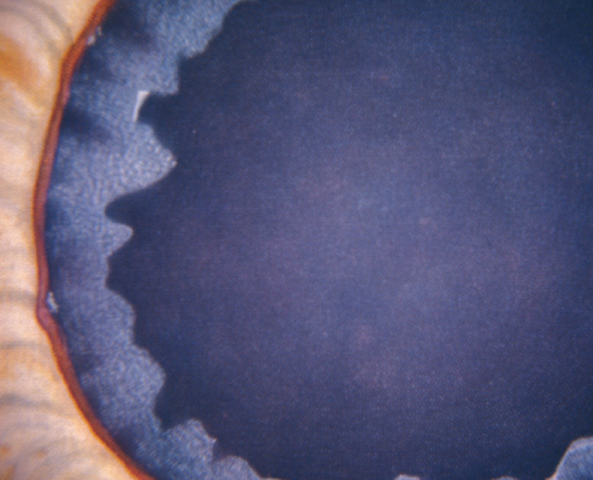
Figure 9.10.1: Pseudoexfoliation syndrome with white material on pupillary margin.
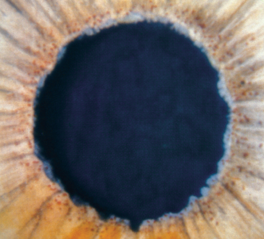
Figure 9.10.2: Pseudoexfoliation syndrome with white material on anterior lens capsule.
Critical
White, flaky material on the pupillary margin; anterior lens capsular changes (central zone of exfoliation material, often with rolled-up edges, middle clear zone, and a peripheral cloudy zone); peripupillary iris TIDs; and glaucomatous optic neuropathy. Bilateral, but often asymmetric.
Other
Irregular dark pigment deposition on the TM more marked inferiorly than superiorly; black scalloped deposition of pigment anterior to Schwalbe line (Sampaolesi line) seen on gonioscopy, especially inferiorly. White, flaky material may be seen on the corneal endothelium, which may have a lower-than-normal endothelial cell density; can look like angular, irregular KP. Iris atrophy. Poor pupillary response to dilation (with more advanced cases, believed to be secondary to iris dilator muscle atrophy). Incidence increases with age. Zonular laxity can lead to anterior lens dislocation, angle narrowing, and secondary angle closure.
Every 1 to 3 months as with POAG, but with the awareness that damage can progress very rapidly.
|
 NOTE NOTEMany patients have pseudoexfoliation syndrome without glaucoma. These patients are reexamined every 6 to 12 months because of glaucoma risk; treatment is initiated with evidence of IOP elevation and glaucomatous damage. |