Severe and boring eye pain (most prominent feature), which may radiate to the forehead, brow, jaw, or sinuses and classically awakens the patient at night. Pain worsens with eye movement and with touch. Gradual or acute onset with red eye. May have tearing, photophobia, or a decrease in vision. Recurrent episodes are common. Scleromalacia perforans (necrotizing scleritis without inflammation) may have minimal symptoms.
Inflammation of scleral, episcleral, and conjunctival vessels (scleral vessels are large, deep vessels that cannot be moved with a cotton swab and do not blanch with topical 2.5% or 10% phenylephrine). Can be sectoral, nodular, or diffuse with associated scleral edema. Characteristic violaceous scleral hue (best seen in natural light by gross inspection). Areas of scleral thinning or remodeling may appear with recurrent episodes, allowing the underlying uvea to become visible or even bulge outward.
Scleral nodules, corneal changes (peripheral keratitis, limbal guttering, or keratolysis), glaucoma, uveitis, or cataract.
Subretinal granuloma, circumscribed fundus mass, choroidal folds, retinal striae, exudative retinal detachment, optic disc swelling, macular edema, proptosis, or rapid-onset hyperopia.
Episcleritis: Sclera not involved. Blood vessels blanch with topical phenylephrine. Usually more acute onset than scleritis. Patients tend to be younger and have mild symptoms, if any. See 5.6, Episcleritis.
VogtKoyanagiHarada (VKH) disease, choroidal melanoma, metastatic choroidal tumor, and choroidal hemangioma can mimic posterior scleritis.
Scleritis associated with other orbital inflammatory foci (myositis, dacryoadenitis, etc.) may be part of idiopathic orbital inflammatory syndrome (IOIS) commonly known as orbital pseudotumor.
Up to 50% of patients with scleritis have an associated systemic disease, typically connective tissue or vasculitic in nature. Workup indicated if no known underlying disease is present.
Connective tissue disease (e.g., rheumatoid arthritis, granulomatosis with polyangiitis, relapsing polychondritis, systemic lupus erythematosus, reactive arthritis, polyarteritis nodosa, ankylosing spondylitis, inflammatory bowel disease), infectious (e.g., Pseudomonas, atypical mycobacteria, fungi, Nocardia, syphilis), trauma including status-post surgery (especially scleral buckle or pterygium surgery with mitomycin-C or beta irradiation), and gout.
Varicella zoster, tuberculosis, Lyme disease, other bacteria (e.g., Pseudomonas species with scleral ulceration, Proteus species associated with scleral buckle), sarcoidosis, foreign body, medications (e.g., pembrolizumab), or parasite.
Classification
Diffuse anterior scleritis: Widespread inflammation of the anterior segment.
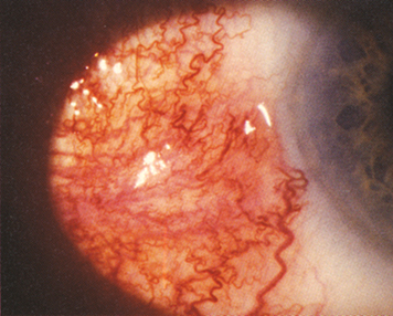
Nodular anterior scleritis: Immovable inflamed nodule(s) (see Figure 5.7.1).
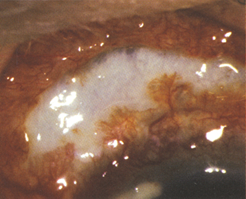
Necrotizing anterior scleritis with inflammation (see Figure 5.7.2): Extreme pain. The sclera becomes transparent (choroidal pigment visible) because of necrosis. High association with systemic inflammatory diseases.
Necrotizing anterior scleritis without inflammation (scleromalacia perforans): Typically asymptomatic. Seen most often in older women with long-standing rheumatoid arthritis.
Posterior scleritis: May start posteriorly, or rarely be an extension of anterior scleritis, or simulate an amelanotic choroidal mass. Associated with exudative retinal detachment, disc swelling, retinal hemorrhage, choroidal folds, choroidal detachment, restricted motility, proptosis, pain, or tenderness.
History: Previous episodes? History of ocular trauma or surgery? Medical problems? An associated systemic disease is more common in patients >50 years old.
Examine the sclera in all directions of gaze by gross inspection in natural light or adequate room light.
Slit-lamp examination with a red-free filter (green light) to determine whether avascular areas of the sclera exist. Check for corneal or anterior chamber involvement.
Dilated fundus examination to rule out posterior involvement.
B-scan ultrasound to detect posterior scleritis (e.g., T-sign).
Fluorescein angiography in eyes with posterior scleritis may show multiple areas of pinpoint leakage, choroidal folds, and subretinal fluid.
Complete physical examination (especially joints, skin, and cardiovascular and respiratory systems) by an internist or a rheumatologist.
Complete blood count, creatinine, ESR, C-reactive protein (CRP), uric acid, RPR or VDRL, FTA-ABS or MHA-TP, ANCA, rheumatoid factor, anti-CCP, ANA, ACE, CH50 (total complement activity assay), C3, C4, and urinalysis.
Other tests if clinical suspicion warrants additional workup: PPD or IGRA, Lyme antibody, chest x-ray, HLA B27, x-ray of sacroiliac joints, and MRI or CT scan if indicated. Cultures should be taken if infection is suspected.
Diffuse and nodular scleritis: One or more of the following may be required. Concurrent antiulcer medication (e.g., proton-pump inhibitor [e.g., omeprazole 20 mg p.o. daily] or histamine type 2 receptor blocker [e.g., ranitidine 150 mg p.o. b.i.d.]) may be helpful.
Oral NSAIDs (e.g., flurbiprofen 100 mg t.i.d., naproxen 250 to 500 mg p.o. b.i.d., or indomethacin 25 to 50 mg p.o. t.i.d.): Several different NSAIDs may be tried before therapy is considered a failure. If there is still no improvement, consider systemic steroids.
Oral steroids: Prednisone 60 to 80 mg p.o. daily for 1 week, followed by a taper to 20 mg daily over the next 2 to 6 weeks, followed by a slower taper. Once daily calcium with vitamin D (e.g., 600 mg with 400 iU) supplements should be given to reduce the risk of osteoporosis. An oral NSAID often facilitates the tapering of the steroid but significantly increases the risk of gastric ulceration. If unsuccessful or disease requires >7.5 to 10 mg prednisone/d for long-term control, immunosuppressive therapy is indicated.
Intravenous steroids: Methylprednisolone succinate 1,000 mg daily for 3 days (followed by oral steroids as above) is preferable to prednisone >80 mg/d because of reduced risk of ischemic necrosis of bone.
Immunosuppressive therapy (e.g., cyclophosphamide, methotrexate, cyclosporine, azathioprine, mycophenolate mofetil, anti-tumor necrosis factor alpha (TNFα) agents, and other biologics): If one drug is ineffective or not tolerated, additional agents should be tried. Systemic steroids may be used in conjunction. Immunosuppressive therapy should be coordinated with an internist, rheumatologist, or uveitis specialist. Topical cyclosporine is rarely effective.
Conventional teaching is that topical therapy is of little benefit. However, difluprednate 0.05% drops are sometimes helpful (with or without a topical NSAID) and thus, in mild cases, may spare the need for systemic immunosuppressive agents.
Subconjunctival steroid injections (e.g., 0.1 to 0.3 mL of triamcinolone acetonide 40 mg/mL or dexamethasone sodium phosphate 4 mg/mL): May be very helpful in patients unable to tolerate systemic therapy. Side effects may include subconjunctival hemorrhage, cataract, glaucoma, and (rarely) catastrophic scleral melting. Do not use in cases of necrotizing scleritis.
Necrotizing scleritis associated with rheumatoid arthritis is associated with increased mortality due to coronary arteritis or cerebral angiitis and requires urgent, aggressive immunosuppressive therapy.
Systemic steroids and immunosuppressive therapies are used as above during the first month; the former is tapered slowly.
Scleral patch grafting may be necessary if there is a significant risk of perforation, ideally once the inflammation is better controlled.
Posterior scleritis: Therapy may include systemic aspirin, NSAIDs, steroids, or immunosuppressive therapy as described previously. Consult a retina or uveitis specialist.
Infectious etiologies: Debridement and cultures/stains are essential. Treat with appropriate topical and systemic antimicrobials. Oral fluoroquinolones have good ocular tissue penetration. If a foreign body (e.g., scleral buckle [associated with Proteus or Pseudomonas]) is present, surgical removal is indicated.
Glasses or eye shield should be worn at all times if there is significant thinning and risk of perforation.