section name header

Symptoms

Acute or rapid onset of redness and mild pain in one or both eyes, typically in young to middle-aged adults, more common in women; a history of recurrent episodes is common. No discharge or photophobia.

Signs

Critical

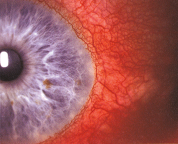
Sectoral (and, less commonly, diffuse) redness of one or both eyes, mostly due to engorgement of the episcleral vessels. These vessels are large, run in a radial direction beneath the conjunctiva, and can be moved slightly with a cotton-tip applicator (see Figure 5.6.1).

Figure 5.6.1: Episcleritis.

Rapuano9781975243722-ch005_f014.jpg

Other

Mild-to-moderate tenderness over the area of episcleral injection or a nodule that can be moved slightly over the underlying sclera may be seen. Fluorescein staining can sometimes be seen over the nodule. Associated anterior uveitis and corneal involvement are rare. Vision is normal.

Differential Diagnosis

Etiology

Workup

  1. History: Assess for a history of rash, arthritis, venereal disease, recent viral illness, and other medical problems.

  2. External examination in natural light: Look for the violaceous hue of scleritis.

  3. Slit-lamp examination: Anesthetize (e.g., topical proparacaine) and move the conjunctiva with a cotton-tipped applicator to determine the depth of the injected blood vessels. Evaluate for any corneal or anterior chamber involvement. Check IOP.

  4. Place a drop of phenylephrine 2.5% in the affected eye and re-examine the vascular pattern 10 to 15 minutes later. Episcleral vessels should blanch, highlighting any underlying scleral vascular engorgement.

  5. If the history suggests an underlying etiology (systemic disease usually precedes ocular involvement), or in cases with multiple recurrences, the appropriate laboratory tests should be obtained (e.g., complete blood count, comprehensive metabolic panel, antinuclear antibody [ANA], rheumatoid factor, anticyclic citrullinated peptide [anti-CCP], erythrocyte sedimentation rate [ESR], serum uric acid level, RPR or VDRL, FTA-ABS or MHA-TP, antineutrophil cytoplasmic antibody [ANCA]).

Treatment

  1. If mild, treat with artificial tears q.i.d.

  2. If moderate to severe, a topical NSAID (e.g., diclofenac 0.1% q.i.d., bromfenac 0.07% or 0.09% daily) or a mild topical steroid (e.g., fluorometholone 0.1% or 0.25%, fluorometholone acetate 0.1%, or loteprednol 0.5% q.i.d.) often relieves the discomfort. Occasionally, more potent or frequent topical steroid application is necessary.

  3. Oral NSAIDs may be used as an alternate steroid-sparing initial therapy and should be given with food or antacids (e.g., ibuprofen 200 to 600 mg p.o. t.i.d. to q.i.d., naproxen 250 to 500 mg p.o. b.i.d., or flurbiprofen 50 to 100 mg p.o. b.i.d. to t.i.d.) for at least 10 to 14 days.

NOTE

Many physicians prefer oral NSAIDs to topical NSAIDs or steroids as initial therapy.

Follow-Up

Patients treated with artificial tears need not be seen for several weeks unless discomfort worsens or persists. If topical steroids are used, recheck every 2 to 3 weeks until symptoms resolve. The frequency of steroid administration is then tapered. Episcleritis may recur in the same or contralateral eye.