Acute: Pain, redness, photophobia, consensual photophobia (pain in the affected eye when a light is shone in the fellow eye), excessive tearing, and decreased vision.
Chronic: Decreased vision (from cataract, vitreous debris, CME, epiretinal membrane [ERM]), and/or floaters. May have periods of exacerbations and remissions with few acute symptoms (e.g., juvenile idiopathic arthritis [JIA]associated uveitis).
Cells and flare in the AC (see Tables 12.1.1 and 12.1.2) and ciliary flush.
Stellate KPsmall KP with multiple fine projections from their core, appearing star-shaped.
Granulomatous KPlarge KP with a greasy or mutton fat appearance.
Layering of white blood cells in the AC.
Differential for hypopyon uveitis includes HLA-B27, sarcoidosis, phacogenic anaphylaxis, endophthalmitis, syphilis, TB, Behçet disease, herpetic, rifabutin, and the pseudohypopyon of lymphoma, leukemia, retinoblastoma, or triamcinolone.
A shifting hypopyon (moves with head position) suggests Behçet disease, lymphoma, or pseudohypopyon from prior triamcinolone injection.
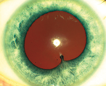
Low intraocular pressure (IOP) (more common, secondary to ciliary body hyposecretion), elevated IOP (e.g., herpetic, lens induced, FHIC, PosnerSchlossman syndrome), fibrin (e.g., HLA-B27, endophthalmitis), iris nodules (e.g., sarcoidosis, syphilis, TB), iris atrophy (e.g., herpetic, fourth-generation fluoroquinolones), iris heterochromia (e.g., FHIC), iris synechiae (especially HLA-B27, sarcoidosis), band keratopathy (especially JIA in younger patients, any chronic uveitis in older patients), uveitis in a quiet eye (consider JIA, FHIC, and masquerade syndromes), and CME (see Figure 12.1.1).
Diagnostic Categories
Tight (contact) lens syndrome. See 4.21, Contact Lens-Related Problems.
Neoplasms: Pseudohypopyon of retinoblastoma (in children), leukemia, and lymphoma; recurrent hyphema of juvenile xanthogranuloma (in children), metastatic disease.
Ocular ischemic syndrome (OIS). See 11.11, Ocular Ischemic Syndrome/Carotid Occlusive Disease.
Pigmentary dispersion syndrome. See 9.9, Pigment Dispersion Syndrome/Pigmentary Glaucoma.
SchwartzMatsuo syndromepigmentary cells released from a chronic retinal detachment move into AC and trabecular meshwork causing elevated IOP.
Behçet disease: Classic triad of painful oral ulcers, genital ulcers, and uveitis. A skin rash is also common (pathergyformation of a pustule on skin after needle puncture). Hypopyon uveitis is found in 5% to 15% of patients with uveitis. May be associated with retinal vasculitis which can be occlusive and result in profound vision loss. See 12.5, Retinal Vasculitis. In its most severe form it can cause a panuveitis. See 12.4, Panuveitis.
Brimonidine-associated uveitis: Occurs in conjunction with brimonidine-associated allergic papillary conjunctivitis. Granulomatous KP are out of proportion to AC cell.
CMV-associated anterior uveitis: May be chronic or acute and recurrent. Can have associated elevated IOP with flares similar to HSV/VZV. More common in those of Asian ancestry. Often have characteristic coin-shaped KP not seen in other herpetic uveitis. May require long-term oral valganciclovir and topical steroids for chronic suppression.
FHIC: Patients mainly complain of floaters with few other symptoms. There are characteristic stellate KP distributed diffusely over the corneal endothelium and are out of proportion to AC cell, diffuse iris stromal atrophy causing a lighter appearing iris color and transillumination defects. Gonioscopy may reveal fine vessels that cross the trabecular meshwork. Vitreous opacities are common. Glaucoma and cataract are also common, typically secondary to (inappropriate) corticosteroid use. Corticosteroids are not helpful. CME and posterior synechiae are absent. Cataract surgery may cause AC hemorrhage from rupture of fine angle vessels (Amsler sign), but outcomes are usually excellent.
HLA-B27: Explosive in onset. Typically alternates eyes, but 10% are simultaneous bilateral. Systemic manifestations including ankylosing spondylitis, reactive arthritis (previously Reiter syndrome), psoriatic arthritis, and inflammatory bowel disease (IBD).
Bilateral acute recurrent alternating anterior uveitis is very characteristic of HLA-B27 uveitis. |
HSV/VZV-associated: May be chronic or acute and recurrent. May have associated elevated IOP with flares. May have associated periocular rash. May have associated corneal disease. See 4.16, Herpes Simplex Keratitis and 4.17, Herpes Zoster Ophthalmicus/Varicella Zoster Virus. Sectoral transillumination defects are common in VZV while more diffuse defects are seen in HSV. Care should be taken to rule out associated retinitis. See 12.13, Acute Retinal Necrosis.
JIA-associated: Most common in females, oligoarthritis (≤4 joints involved) that starts before age 4 years, antinuclear antibody (ANA) positive, and rheumatoid factor (RF) negative. May be associated with glaucoma, cataracts, band keratopathy, and CME. Uveitis occurs less commonly in polyarticular and rarely in systemic JIA (Still disease).
Phacogenic anaphylaxis: Immune reaction to lens material, often secondary to aborted or incomplete cataract extraction, trauma with lens capsule violation, or hypermature cataract. See 9.11, Lens-Related Glaucoma.
PosnerSchlossman syndrome: Recurrent episodes of very high IOP and mild AC inflammation. Many cases are caused by herpetic uveitis (HSV, VZV, or CMV). See 9.7, Uveitic Glaucoma.
PUPPI: Chronic AC inflammation following intraocular surgery. Rule out acute endophthalmitis, retained lens fragments, UGH syndrome, or recurrence of preexisting anterior uveitis (e.g., HLA-B27associated uveitis). A small percentage of patients with well-positioned posterior chamber intraocular lenses (IOL) may develop a persistent low-grade, steroid-responsive anterior uveitis that recurs when low-dose topical steroids are tapered off. Postoperative endophthalmitis must be considered. See 12.15, Subacute and Chronic Postoperative Uveitis.
Sarcoidosis: Pleomorphic uveitis. May be unilateral, bilateral, acute, chronic, granulomatous, or nongranulomatous. More common in those of African or Scandinavian descent. The most common systemic manifestation in adults is within the lungs and a rash in children. Unique anterior segment findings of sarcoidosis include iris and conjunctival granulomas (the latter can be biopsied to help make a diagnosis).
Syphilis: See 12.10, Syphilis.
Traumatic iritis: See 3.6, Traumatic Iritis.
TINU: Uncommon but frequently underdiagnosed; classically a chronic bilateral nongranulomatous uveitis in children and young adults with a female predilection. May be precipitated by oral nonsteroidal anti-inflammatory drug (NSAID) therapy. Systemic symptoms include abdominal pain, fatigue, and malaise.
UGH syndrome: Secondary to IOL-iris chafe resulting in recurrent or chronic inflammation, hyphema, and elevated IOP. More common with sulcus-based IOLs. Assess closely for location of the IOL, pseudophacodonesis, and for IOL subluxation. Transillumination defects are common in the area of IOL-iris touch. See 9.15, Postoperative Glaucoma.
Complete ocular examination, including an IOP check, gonioscopy, and a dilated fundus examination. The vitreous and fundus should be evaluated for signs of intermediate and posterior uveitis.
A diagnostic workup may be unnecessary in the setting of a first episode of a mild, unilateral, nongranulomatous uveitis with a history and examination that are not suggestive of associated systemic disease.
In most other situations, a targeted workup is recommended. If too many tests are ordered unnecessarily, a portion of them may come back falsely positive and shroud the diagnosis. See Table 12.1.3. However, if a patient presents with bilateral, granulomatous, or recurrent anterior uveitis the patient should be evaluated for sarcoidosis, syphilis, TB (in at-risk patients), and additional workup should be focused based on history and examination.
Treponemal test (syphilis enzyme immunoassay [EIA], fluorescent treponemal antibody absorption [FTA-ABS], Treponema pallidum particle agglutination [TP-PA]), followed by confirmatory nontreponemal test (rapid plasma reagin [RPR], venereal disease research laboratory [VDRL]). See 12.10, Syphilis.
Interferon gamma releasing assay (IGRA, QuantiFERON Gold) and/or purified protein derivative (PPD). Consider chest imaging (e.g., chest x-ray [CXR] or computed tomography [CT] chest) to assess for signs of active or prior pulmonary disease.
Serologic testing for antibodies against HSV, VZV, CMV, and toxoplasmosis can be useful in ruling out a disease as the causative etiology (IgG and/or IgM negative). The presence of positive IgG titer is not necessarily indicative of active disease, but rather prior exposure. Very elevated IgG titers may make you more suspicious for active disease, and a positive IgM indicates recent infection, but again are not fully diagnostic. Negative testing helps to rule out the disease.
AC paracentesis for polymerase chain reaction (PCR) testing for suspected herpes virusassociated anterior uveitis (HSV, VZV, and CMV).
Angiotensin-converting enzyme (ACE), lysozyme, and chest imaging (CXR or CT chest).
HLA-B27 antigen typing (in acute unilateral or bilateral alternating anterior uveitis; especially if hypopyon is present; ask about symptoms of spondyloarthropathy, psoriasis, and reactive arthritis).
HLA-B51 testing is neither sensitive nor specific and has minimal clinical utility. Behçet disease is a clinical diagnosis.
Urinary beta-2 microglobulin, urinalysis, and renal function testing are useful in making a diagnosis of TINU.
TABLE 12.1.3: Suggested Diagnostic Workup for Anterior Uveitis
| Suspected Etiology | Suggested Workup |
|---|---|
| Ankylosing spondylitis | HLA-B27, SI joint films, rheumatology consult |
| HSV/VZV/CMV | AC tap to detect HSV/VZV/CMV DNA via PCR; consider HSV serologies, VZV serologies, and CMV serum PCR testing. |
| Juvenile idiopathic arthritis | Rheumatoid factor, antinuclear antibodies, HLA-B27, radiographs of affected joints, urinalysis, and renal function testing; pediatric rheumatology consult |
| Ocular ischemic syndrome | Intravenous fluorescein angiography, carotid Doppler studies |
| Psoriatic arthritis | HLA-B27; rheumatology and/or dermatology consult |
| Reactive arthritis | HLA-B27, SI joint films (if symptomatic); referral for gastrointestinal or genitourinary inciting infection (campylobacter, chlamydia, salmonella, shigella). |
| Sarcoidosis | CXR and/or CT chest, PPD or IGRA, ACE, lysozyme |
| Syphilis | RPR or VDRL, treponemal test (FTA-ABS, TPPA, EIA) followed by nontreponemal test (RPR, VDRL); FTA-ABS or treponemal-specific assay; HIV testing if positive |
| TINU | Urine beta-2 microglobulin, urinalysis, renal function testing; nephrology consultation |
Cycloplegic (e.g., cyclopentolate 1% b.i.d. to help prevent posterior synechiae formation; cyclopentolate 1% t.i.d. or atropine 1% b.i.d. to help break early formed posterior synechiae).
Topical steroid (e.g., prednisolone acetate 1%) q16h, depending on the severity of inflammation. Most cases of moderate to severe acute uveitis require q12h dosing initially. Difluprednate 0.05% may allow less frequent dosing than prednisolone acetate. Consider a loading dose (prednisolone acetate 1% one drop every minute for 5 minutes at bedtime and awakening) or fluorometholone 0.1% ophthalmic ointment at night.
If the uveitis is severe and is incompletely responsive to topical steroids, consider a course of oral steroids (after ruling out infectious etiologies).
If the disease becomes persistent (lasting >3 months) or is frequently recurrent (≥3 episodes per year) consider consultation with a rheumatologist or uveitis specialist for possible steroid-sparing immunosuppressive therapy.
Treat secondary glaucoma with aqueous suppressants. Avoid pilocarpine. Glaucoma may result from:
Cellular blockage of the trabecular meshwork. See 9.7, Uveitic Glaucoma.
Secondary angle closure from synechiae formation. See 9.4, Acute Angle Closure Attack and 9.5, Chronic Angle Closure Glaucoma.
Neovascularization of the iris and angle. See 9.13, Neovascular Glaucoma.
Steroid response. See 9.8, Steroid-Response Glaucoma.
Although topical prostaglandins may rarely cause uveitis and CME, they can be tried if other medical management is ineffective before considering glaucoma surgery.
If an exact etiology for the anterior uveitis is determined, then additional ocular and/or systemic management may be indicated.
Behçet disease: Typically requires systemic immunosuppression. Antitumor necrosis factor (TNF) medications and azathioprine are considered first-line agents.
Ankylosing spondylitis: Often requires systemic anti-inflammatory agents (e.g., NSAIDs such as naproxen). Consider consulting rheumatology, physical therapy, and cardiology (increased incidence of cardiomegaly, conduction defects, and aortic insufficiency).
IBD: Often benefits from systemic steroids, sulfadiazine, or other immunosuppressive agents. Obtain a medical or gastrointestinal consult.
Psoriatic arthritis: Consider a rheumatology and/or dermatology consult.
Reactive arthritis: If urethritis is present, then the patient and sexual partners are treated for chlamydia (e.g., doxycycline 100 mg b.i.d. for 7 days or alternatively a single-dose azithromycin 1 g p.o.). Obtain medical and/or rheumatology or urology consult.
FHIC: Usually does not respond to nor require steroids (a trial of steroids may be attempted, but they should be tapered quickly if there is no response); cycloplegics are not necessary.
PosnerSchlossman syndrome: See 9.7, Uveitic Glaucoma.
Herpetic uveitis: Herpetic iridocyclitis benefits from topical steroids and systemic antiviral medications; topical antivirals are usually ineffective for uveitis due to poor intraocular penetration. See 4.15, Herpes Simplex Virus and 4.16, Herpes Zoster Ophthalmicus/Varicella Zoster Virus.
JIA-associated uveitis: Topical steroids can be useful acutely for reducing cells and flare, but should be minimized for long-term therapy to reduce the risk of cataract and glaucoma, both of which are more common in children. Systemic steroid therapy in children may cause growth suppression and should be avoided if possible. Prolonged cycloplegic therapy may be required and necessitate appropriate refractive correction. Consultation with rheumatology, pediatrics, and/or a uveitis specialist is useful as immunomodulatory therapy (e.g., methotrexate, adalimumab) is often needed. Regular follow-up is essential, as flares may be asymptomatic; recurrent or chronic disease can lead to irreversible damage and various sequelae including synechiae, glaucoma (or hypotony), CME, ERM, and cataract formation.
Phacoantigenic glaucoma: Usually requires removal of the lens material. See 9.11, Lens-Related Glaucoma.
Sarcoidosis: Refer patients to an internist or pulmonologist for systemic evaluation and medical management which may include systemic steroids and immunosuppressives. Consider early referral to a uveitis specialist in complicated cases. A poor visual outcome has been reported with posterior uveitis, glaucoma, delay in definitive treatment, or presence of macula-threatening conditions such as CME.
Syphilis: See 12.10, Syphilis.
Traumatic iritis: See 3.6, Traumatic Iritis.
TINU: Low-dose topical steroids are often enough to suppress mild, chronic anterior segment inflammation. If there is renal involvement, consultation with a nephrologist is necessary to consider systemic immunosuppression to prevent kidney damage.
TB: Refer the patient to an internist, infectious disease specialist, or public health officer for consideration of systemic treatment. Patients with ocular TB frequently have no pulmonary disease but still require systemic four-drug antituberculous therapy. Concomitant oral steroids or steroid-sparing immunosuppression may be necessary.
UGH syndrome: See 9.15, Postoperative Glaucoma.
Every 1 to 7 days in the acute phase, depending on the severity; every 1 to 3 months when stable.
A vitreous and fundus examination should be performed with all flare-ups, decreased vision, or every 3 to 6 months. Macular edema is a frequent cause of decreased vision even after the uveitis is controlled; OCT of the macula can be very useful.
If the AC reaction has resolved, then the steroid drops can be slowly tapered with intermittent examinations to ensure that the inflammation does not recur during the taper (usually by one drop per day every 7 to 14 days [e.g., q.i.d. for 1 week, then t.i.d. for 1 week, then b.i.d. for 1 week, etc.]). Steroids are usually discontinued following the taper when the AC is quiet. Occasionally, long-term, low-dose steroids (q.o.d. to b.i.d.) are required to keep the inflammation from recurring. Punctal occlusion techniques may increase the potency of the drug and decrease systemic absorption. The cycloplegic agents can be discontinued after the AC reaction has resolved.