section name header

General Information

Definition

Glaucoma caused by a fibrovascular membrane overgrowing the anterior chamber angle structures. Initially, despite an open appearance on gonioscopy, the angle may be blocked by the membrane. The fibrovascular membrane eventually contracts, causing PAS formation and secondary angle-closure glaucoma. Rarely, it may have NV of the angle without NV of the iris (NVI) at the pupillary margin. Ischemia-driven vascular endothelial growth factor (VEGF) released from a variety of causes results in the formation of the fibrovascular membrane.

Symptoms

May be asymptomatic or include pain, redness, photophobia, and decreased vision.

Signs

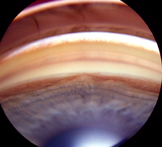
(See Figures 9.13.1 and 9.13.2.)

Figure 9.13.1: Iris neovascularization.

Rapuano9781975243722-ch009_f013.jpg

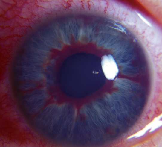
Figure 9.13.2: Neovascularization of the angle.

Rapuano9781975243722-ch009_f014.jpg

Critical

Other

Mild anterior chamber cell and flare, conjunctival injection, corneal edema with acute IOP increase, hyphema, eversion of pupillary margin allowing visualization of iris pigment epithelium (ectropion uveae), optic nerve cupping, and visual field loss.

Differential Diagnosis

Etiology

Workup

  1. History: Determine underlying etiology.

  2. Complete ocular examination, including IOP measurement and gonioscopy to evaluate degree of angle closure, if any. A dilated retinal examination is essential in determining the etiology and for disc evaluation.

  3. Fluorescein angiography as needed to identify an underlying retinal abnormality or in preparation for panretinal photocoagulation (PRP).

  4. Carotid Doppler studies to rule out stenosis when no retinal etiology is identified.

  5. B-scan US or UBM as indicated when the retina cannot be visualized to rule out an intraocular tumor or retinal detachment.

Treatment

  1. Reduce inflammation and pain: Topical steroid (e.g., prednisolone acetate 1% q1–6h) and a cycloplegic (e.g., atropine 1% t.i.d.).

  2. Reduce the IOP if it is increased (markedly high IOP is not uncommon). Where visual potential is good and cupping advanced, a lower target IOP may be appropriate. Any or all of the following medications are used:

    • Topical β-blocker (e.g., timolol 0.5% daily or b.i.d.).

    • Topical α2 agonist (e.g., brimonidine 0.1% to 0.2% b.i.d. to t.i.d.).

    • Topical and/or systemic CAI (e.g., dorzolamide 2% b.i.d. to t.i.d. and/or acetazolamide 500 mg sequel p.o. b.i.d.).

    • Topical rho kinase inhibitor (e.g., netarsudil 0.02% q.d.).

    • Prostaglandins may help lower IOP, but may increase inflammation and are usually avoided in the acute phase.

    • If need for IOP reduction is urgent or refractory to therapies listed above, consider an osmotic agent (e.g., mannitol 1 to 2 g/kg i.v. over 45 minutes).

      NOTE

      Miotics (e.g., pilocarpine) are contraindicated because of their effects on the blood–aqueous barrier. Epinephrine compounds (e.g., dipivefrin) are usually ineffective.

  3. In the acute stages, after a rapid rise in IOP, an anterior chamber paracentesis may be helpful. Caution should be exercised as this may result in a hyphema. See Appendix 13, Anterior Chamber Paracentesis.

  4. If retinal ischemia is thought to be responsible for the NV, then treat with PRP and/or anti-VEGF intravitreal injections. If the retina cannot be visualized, lower the IOP and treat the retina once the cornea clears. These procedures are used if the angle is open as it may be possible to reverse the angle NV and restore normal aqueous outflow.

  5. Glaucoma filtration surgery may be performed when the NV is inactive and the IOP cannot be controlled with medical therapy. Tube-shunt procedures may be helpful to control IOP in some patients with active NV, but may be complicated by postoperative bleeding.  They should not be performed unless there is useful vision to preserve. Transscleral cyclophotocoagulation is an option but is more often reserved for cases with poor visual potential.

  6. Intravitreal anti-VEGF agents (e.g., ranibizumab, bevacizumab, or aflibercept) may be used to promote regression of iris NV prior to, or in conjunction with, filtering surgery or PRP. Their effect is temporary, and their use for treatment of NV is currently off-label. They are particularly useful in stage 1 and 2 neovascular glaucoma, where the angle is still open, to prevent angle closure during the interval required for PRP to take effect. Caution should be used when no view of the retina is possible (See 11.12, Diabetic Retinopathy and 11.17, Neovascular or Exudative [Wet] Age-Related Macular Degeneration, for a discussion on anti-VEGF agents).

  7. In eyes without useful vision, topical steroids and cycloplegics may be adequate therapy for pain control. The pain in chronic neovascular glaucoma is not primarily a function of the IOP itself; thus, reducing IOP may not be necessary if the goal is pain control and comfort measures only. See 13.10, Blind, Painful Eye.

Follow-Up

The presence of NVI, especially with high IOP, requires urgent therapeutic intervention, usually within 1 to 2 days. Angle closure can proceed rapidly (days to weeks).

NOTE

NVI without glaucoma is managed similarly, but there is no need for pressure-reducing agents unless IOP increases.