section name header

Symptoms

Unilateral, painless, acute vision loss (counting fingers to light perception in 94% of eyes) occurring over seconds; may have a history of transient visual loss (amaurosis fugax).

Signs

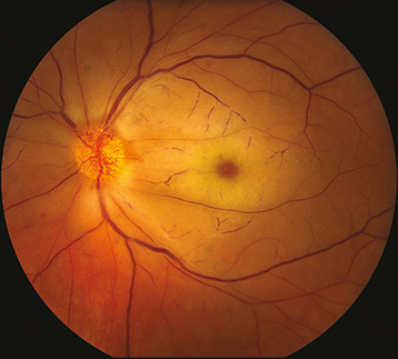
(See Figure 11.6.1.)

Figure 11.6.1: Central retinal artery occlusion.

Rapuano9781975243722-ch011_f012.jpg

Critical

Superficial opacification or whitening of the retina in the posterior pole and a cherry-red spot in the center of the macula (may be subtle).

Other

Marked RAPD. Narrowed retinal arterioles; boxcarring or segmentation of the blood column in the arterioles. Occasionally, retinal arteriolar emboli or cilioretinal artery sparing of the foveola is evident. If visual acuity is light perception or worse, strongly suspect ophthalmic artery occlusion.

Differential Diagnosis

Etiology

Workup

  1. Should be treated as an acute stroke. The American Academy of Ophthalmology (AAO) 2019 guidelines suggest that all these patients should be sent immediately to an emergency department, preferably one with a stroke center, for evaluation and workup.

  2. Immediate ESR, CRP, and platelets to rule out GCA if the patient is 55 years of age or older and no embolus seen on examination. Query GCA review of systems. If the patient’s history and/or laboratories are consistent with GCA, start high-dose systemic steroids. See 10.17, Arteritic Ischemic Optic Neuropathy (Giant Cell Arteritis).

  3. Check the blood pressure.

  4. Other blood tests: Fasting blood sugar and HbA1c, complete blood count (CBC) with differential, prothrombin time/activated partial thromboplastin time (PT/PTT). In patients younger than 50 years or with appropriate risk factors or positive review of systems, consider lipid profile, antinuclear antibody (ANA), rheumatoid factor, syphilis testing (RPR or VDRL and FTA-ABS or treponemal-specific assay), serum protein electrophoresis, hemoglobin electrophoresis, and further evaluation for hypercoagulable state (see above).

  5. Carotid artery evaluation by duplex Doppler US.

  6. Cardiac evaluation with electrocardiography (ECG), echocardiography, and possibly Holter monitoring or bubble study to evaluate for a patent foramen ovale.

  7. OCT can be very helpful in making the diagnosis. Can also consider IVFA, but in obvious cases this should not delay sending the patient to a stroke center. Less commonly ERG is used.

Treatment

CRAO is treated as an acute stroke and immediate referral to an emergency department with a stroke center is warranted. If GCA is suspected, see 10.17, Arteritic Ischemic Optic Neuropathy (Giant Cell Arteritis) for treatment recommendations.

For specific management of ocular signs and symptoms, there are anecdotal reports of improvement after the following treatments, if instituted within 90 to 120 minutes of the occlusive event. None of these treatments have been proven effective in randomized, controlled clinical trials and should not be considered standard of care.

  1. Immediate ocular massage with ophthalmic contact lens (e.g., gonio lens, fundus contact lens) or digital massage.

  2. Anterior chamber paracentesis: See Appendix 13, Anterior Chamber Paracentesis.

  3. IOP reduction with acetazolamide, 500 mg i.v. or two 250-mg tablets p.o. or a topical beta-blocker (e.g., timolol or levobunolol, 0.5% daily or b.i.d.).

  4. Thrombolysis.

Follow-Up

  1. Follow as directed by managing internist and/or neurologist.

  2. Repeat eye examination in 1 to 4 weeks, checking for neovascularization of the iris/disc/angle/retina (NVI/NVD/NVA/NVE), which develops in up to 20% of patients at a mean of 4 weeks after onset. If neovascularization develops, perform panretinal photocoagulation (PRP) with or without an anti-vascular endothelial growth factor (anti-VEGF) agent.