section name header

Symptoms

Weakness or paralysis of one side of the face, inability to close one eye, excessive drooling.

Signs

(See Figures 10.9.1 and 10.9.2.)

Figure 10.9.1: Isolated peripheral left seventh cranial nerve palsy demonstrating lagophthalmos.

Rapuano9781975243722-ch010_f011.jpg

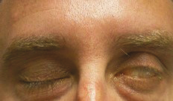
Figure 10.9.2: Isolated peripheral left seventh cranial nerve palsy demonstrating paralysis of upper facial muscles.

Rapuano9781975243722-ch010_f012.jpg

Critical

Unilateral weakness or paralysis of the facial musculature.

Other

Flattened nasolabial fold, droop of corner of the mouth, ectropion, and lagophthalmos. May have ipsilateral decreased taste on anterior two-thirds of tongue, decreased basic tear production, or hyperacusis. May have an injected eye with a corneal epithelial defect. Synkinesis, a simultaneous movement of muscles supplied by different branches of the facial nerve or simultaneous stimulation of visceral efferent fibers of facial nerve (e.g., corner of mouth contracts when eye closes, excessive lacrimation when eating [“crocodile” tears]), secondary to aberrant regeneration implying chronicity.

Etiology

Central Lesions

Peripheral Lesions

Workup

  1. History: Onset and duration of facial weakness? First episode or recurrence? Facial or ear pain? Trauma? Stroke? Recent infection? Hearing loss, tinnitus, dizziness, or vertigo? History of sarcoidosis or cancer?

  2. Examine old photographs to determine chronicity of facial droop.

  3. Complete neurologic examination: Determine whether facial palsy is central or peripheral, complete or incomplete. Look for motor weakness and cerebellar signs. Carefully assess other cranial nerves, especially the fifth, sixth, and seventh. Consider assessing taste on anterior two-thirds of the tongue on affected side.

  4. Complete ocular examination: Check ocular motility and look for nystagmus. Assess orbicularis strength bilaterally, degree of ectropion, and Bell phenomenon. Examine cornea carefully for signs of exposure (superficial punctate keratopathy, epithelial defect, or ulcer). Perform Schirmer test (see 4.3, Dry Eye Syndrome) to assess basic tear production. Check for signs of uveitis.

  5. Otolaryngologic examination: Examine ear and oropharynx for vesicles, masses, or other lesions. Palpate parotid for mass or lymphadenopathy. Check hearing.

  6. CT scan if history of trauma to rule out basilar skull fracture: Axial, coronal, and parasagittal cuts with attention to temporal bone.

  7. MRI or CT scan of the brain if any other associated neurologic signs, history of cancer, or duration >3 months. Sixth cranial nerve involvement warrants attention to the brainstem. Eighth cranial nerve involvement warrants attention to the CPA. Multiple cranial nerve involvement warrants attention to the skull base and cavernous sinus.

  8. CT chest or chest x-ray and angiotensin-converting enzyme (ACE) level if sarcoidosis suspected.

  9. Consider CBC with differential, Lyme antibody, FTA-ABS or treponemal-specific assay, and RPR or VDRL tests depending on suspected etiology.

  10. Consider rheumatoid factor, ESR, antinuclear antibody (ANA), and antineutrophil cytoplasmic antibody if collagen–vascular disease suspected.

  11. Echocardiogram, Holter monitor, and carotid noninvasive studies in patients with a history of stroke.

  12. LP in patients with history of primary neoplasm to rule out carcinomatous meningitis (may repeat if negative to increase sensitivity).

Treatment

  1. Treat the underlying disease as follows:

    • Stroke: Refer to neurologist.

    • CPA masses, temporal bone fracture, nerve laceration: Refer to neurosurgeon. 

    • Otitis: Refer to otolaryngologist.

    • Ramsay–Hunt syndrome: If seen within 72 hours of onset, start acyclovir 800 mg five times per day for 7 to 10 days or valacyclovir 1 g t.i.d. for 7 to 10 days. Dose adjustment is required for patients with renal disease, and careful evaluation of risks and benefits is necessary for pregnant patients. The addition of corticosteroids may be considered. Refer to an otolaryngologist.

    • Guillain–Barré syndrome: Refer to neurologist. May require urgent hospitalization for rapidly progressive motor weakness or respiratory distress.

    • Lyme disease: Refer to infectious disease specialist. May need LP. Treat with oral doxycycline, penicillin, or intravenous (i.v.) ceftriaxone. See 13.4, Lyme Disease.

    • Sarcoidosis: Treat uveitis if present. Consider brain MRI, LP, or both to rule out CNS involvement; if present, refer to neurologist. Refer to internist for systemic evaluation. May require systemic prednisone for extraocular or CNS disease.

    • Metastatic disease: Refer to oncologist. Systemic chemotherapy, radiation, or both may be required.

  2. Bell palsy: The vast majority patients recover completely with observation alone within 3 to 6 months. Options for treatment include the following:

    • Physical therapy with facial massage and/or electrical stimulation of facial musculature.

    • For new-onset Bell palsy, steroids (e.g., prednisone 60 mg p.o. daily for 7 days, followed by a taper of 5 to 10 mg/d) have been shown to increase the likelihood of facial nerve recovery. Antiviral agents, in combination with prednisone, may be offered to patients although the benefit has not been well established.

  3. The primary ocular complication of facial palsy is corneal exposure, which is managed as follows (also see 4.5, Exposure Keratopathy):

    • Mild exposure keratitis: Artificial tears q.i.d. with lubricating ointment q.h.s.

    • Moderate exposure keratitis: Preservative-free artificial tears, gel or ointment q1–2h or moisture chamber with lubricating ointment during the day; moisture chamber with lubricating ointment or tape tarsorrhaphy q.h.s. Consider a temporary tarsorrhaphy.

    • Severe exposure keratitis: Temporary or permanent tarsorrhaphy. For expected chronic facial palsy, consider eyelid gold weight to facilitate eyelid closure.

Follow-Up

  1. Recheck all patients at 1 and 3 months and more frequently if corneal complications arise.

  2. If not resolved after 3 months, order MRI of the brain to rule out mass lesion.

In nonresolving facial palsy with repeatedly negative workup, consider referral to neurosurgeon or plastic surgeon for facial nerve graft, cranial nerve reanastomosis, or temporalis muscle transposition for patients who desire improved facial motor function.