Asymptomatic if isolated. May notice distortion of straight lines or edges, blurred vision, a blind spot, or micropsia if subretinal/submacular fluid develops.
(See Figure 11.34.1.)
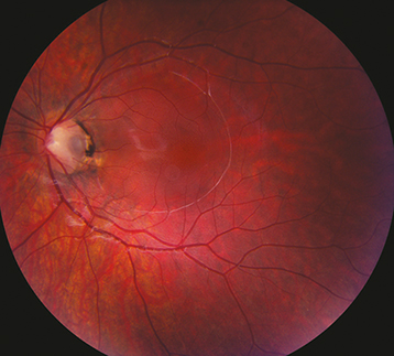
Small, round depression (usually gray, yellow, or black in appearance) in the nerve tissue of the optic disc. Most are temporal, approximately one-third are central, but may be present anywhere on the nerve head.
Peripapillary atrophy, white or gray membrane overlying pit, rarely RAPD, various visual field defects. May develop localized detachment of the sensory retina and/or retinoschisis extending from the disc to the macula, usually unilateral.
Complete ophthalmologic examination including slit-lamp examination of the optic nerve and macula with a handheld lens to evaluate for macular fluid.
Obtain baseline automated visual field testing (e.g., Octopus, Humphrey).
If macular fluid is present, consider OCT to characterize precisely and IVFA to rule out CSCR or CNV.
Isolated optic pits: Yearly examination including IOP check, dilated fundus examination, and visual field testing if indicated; sooner if symptomatic. Patients should be given an Amsler grid. See Appendix 4, Amsler Grid. Monitor for and treat amblyopia if present.
Optic pits with serous macular detachment or retinoschisis: Refer for retinal evaluation.