Author(s): Gregory H.Foos, JeanKwo
- The pharynx is divided into the nasopharynx, the oropharynx, and the laryngopharynx.
- The nasopharynx consists of the nasal passages, including the septum, turbinates, and adenoids.
- The oropharynx consists of the oral cavity, including the dentition and tongue.
- The epiglottis separates the laryngopharynx into the larynx (leading to the trachea) and the hypopharynx (leading to the esophagus).
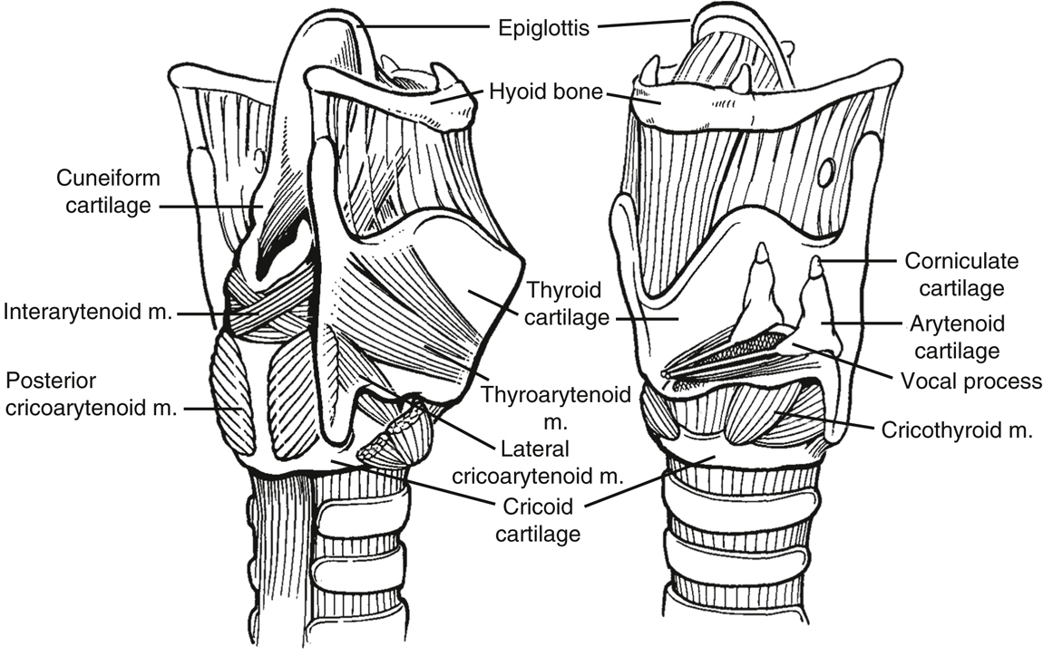
- The larynx (see Figure 13.1)
- The larynx, located at the level of the fourth to the sixth cervical vertebrae, originates at the laryngeal inlet and ends at the inferior border of the cricoid cartilage. It consists of nine cartilages: three unpaired (thyroid, cricoid, and epiglottis) and three paired (corniculates, cuneiforms, and arytenoids); ligaments; and muscles.
- The cricoid cartilage, located just inferior to the thyroid cartilage at the C5-6 vertebral level, is the only complete cartilaginous ring in the respiratory tree. Because it is a complete ring, pressure is applied here (Sellick maneuver) to occlude the esophagus when performing a rapid sequence induction.
- The cricothyroid membrane connects the thyroid and cricoid cartilages and measures 0.9 × 3.0 cm in adults. The membrane is superficial, thin, and devoid of major vessels in the midline, making it an important site for emergent surgical airway access (see Section VI.B.3).
- The laryngeal muscles can be divided into two groups: muscles that open and close the glottis (lateral cricoarytenoid [adduction], posterior cricoarytenoid [abduction], and transverse arytenoid) and muscles that control the tension of the vocal ligaments (cricothyroid, vocalis, and thyroarytenoid).
- Innervation
- Sensory. The glossopharyngeal nerve (cranial nerve IX) provides sensory innervation to the posterior one-third of the tongue, the oropharynx from its junction with the nasopharynx, including the pharyngeal surfaces of the soft palate, epiglottis, and the fauces, to the junction of the pharynx and esophagus. The internal branch of the superior laryngeal nerve, a branch of the vagus nerve (cranial nerve X), provides sensory innervation to the mucosa from the epiglottis to and including the vocal cords. The sensory fibers of the inferior laryngeal nerve, a branch of the recurrent laryngeal nerve (also a branch of the vagus nerve), provide sensory innervation to the mucosa of the subglottic larynx and trachea. A small branch of the glossopharnyngeal nerve, Hering nerve, also serves to transmit information from baroreceptors in the carotid sinus and chemoreceptors in the carotid body to the brainstem.
- Motor. The external branch of the superior laryngeal nerve provides motor innervation to the cricothyroid muscle. Activation of this muscle results in tensing of the vocal cords. The motor fibers of the inferior laryngeal nerve provide motor innervation to all other intrinsic muscles of the larynx. Bilateral injury to the inferior laryngeal nerves (eg, via injury to the recurrent laryngeal nerves) can produce unopposed activation of the cricothyroideus, leading to tensing of the vocal cords and airway closure.
- The glottis is composed of the vocal folds (true and “false” cords) and the rima glottidis.
- The rima glottidis describes the aperture between the true vocal cords.
- The glottis is the narrowest point in the adult airway (more than 8 years of age), whereas the cricoid cartilage is the narrowest point in the infant airway (birth to 1 year of age). Of note, a recent MRI study showed the glottis is the narrowest portion of the sedated, unparalyzed pediatric (2 months-13 years).
- The lower airway extends from the subglottic larynx to the bronchi.
- The subglottic larynx extends from the vocal folds to the inferior border of the cricoid cartilage (C6).
- The trachea is a fibromuscular tube that is 10 to 12 cm long with a diameter of approximately 20 mm in adults. It extends from the cricoid cartilage to the carina. The trachea is supported by 16 to 20 U-shaped cartilages, with the open end facing posteriorly. Noting the posterior absence of cartilaginous rings provides anterior-posterior orientation during fiberoptic exam of the tracheobronchial tree.
- The trachea bifurcates into the right and left main stem bronchi at the carina. The right main stem bronchus is approximately 2.5 cm long with a takeoff angle of approximately 25°. The left main stem bronchus is approximately 5 cm long with a takeoff angle of approximately 45°. Because the right main stem bronchus branches at a less acute angle, aspiration and accidental bronchial intubation are more likely to occur on the right side.
