Molecular mechanism
Propofol is an agonist at postsynaptic gamma-aminobutyric acid type A (GABAA) receptors, inducing an inward chloride current that hyperpolarizes postsynaptic neurons and enhances inhibition.
Neural circuit mechanism
Propofol acts on GABAA receptors at multiple sites, including the cortex, thalamus, brainstem, and spinal cord.
By enhancing GABAergic inhibition in the thalamus and brainstem, which are highly interconnected with the cortex, propofol decreases excitatory inputs and enhances the inhibition of cortical pyramidal neurons.
The potentiation of GABAergic circuits is thought to produce the various features of anesthesia, including loss of consciousness, apnea, and atonia.
EEG signatures
EEG signatures are markers of altered arousal states induced by each anesthetic.
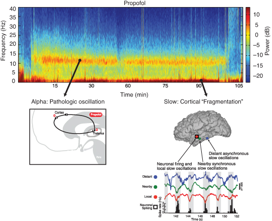
Propofol-induced unconsciousness is characterized by slow–delta (0.1- to 4-Hz) and frontally coherent alpha (8- to 12-Hz) oscillations (Figure 16.5).
FIGURE 16.5 Spectrogram of propofol showing a pathologic alpha oscillation (8 to 12 Hz), which corresponds with prevention of communication between the thalamus and cortex, and slow wave (0.1- to 1-Hz) cortical fragmentation.
EEG analysis showing frequency changes during propofol anesthesia. Includes brain activity graphs, brain diagram with oscillation points, and a schematic of the brain during oscillation.
Slow–delta oscillations are thought to result from GABA-mediated inactivation of arousal circuits and appear to disrupt cortical integration by isolating local cortical networks and impairing intracortical communication. Specifically, slow–delta oscillations may be associated with neurons across the cortex being activated and inactivated at different times (up–down or on–off states). This fragments brain activity into asynchronous windows and impairs communication, which is also referred to as “cortical fragmentation” (Figure 16.5).
Based on modeling studies, alpha oscillations are thought to reflect highly structured rhythms within thalamocortical circuits. During propofol-induced unconsciousness, these abnormally coherent or synchronous alpha oscillations are thought to interfere with communication between the thalamus and frontal cortex (Figure 16.5).
High concentrations of propofol (and several other anesthetics) can lead to burst suppression, which is characterized by alternating periods of brief bursts of high-amplitude activity and isoelectricity (suppression). Intraoperative burst suppression is generally avoided for routine cases owing to concerns about potential adverse effects but can be at times targeted for specific indications like status epilepticus, intracranial hypertension, or deep hypothermic circulatory arrest.
Systematic changes in the amplitude of alpha and frequency of slow–delta oscillations can be used to track anesthetic depth prior to burst suppression. Specifically, as anesthetic concentrations increase, the amplitude of alpha oscillations tends to decrease, and the dominant frequency of slow–delta oscillations tend to increase. Monitoring these changes can help clinicians anticipate a deepening state of unconsciousness before the onset of burst suppression.
Ether-derived anesthetics (sevoflurane, isoflurane, desflurane)
Molecular mechanism
Ether-derived anesthetics bind at multiple targets within the central nervous system, including GABAA receptors, N-methyl-d-aspartate (NMDA) receptors, potassium channels, and others.
Neural circuit mechanism
Although the neural circuit mechanisms of ether-derived anesthetics have not been fully characterized, the EEG signature under general anesthesia with inhaled anesthetics is very similar to propofol-induced anesthesia, suggesting that the primary mechanism is GABAA-mediated inhibition of similar thalamocortical and corticocortical dynamics.
EEG signatures
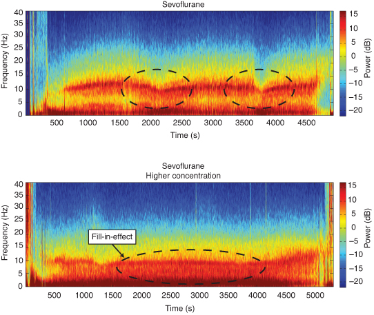
Under sevoflurane general anesthesia, the EEG is characterized by prominent alpha and slow–delta oscillations, similar to propofol general anesthesia, with the addition of theta (4- to 8-Hz) oscillations, particularly at higher concentrations.
The addition of theta oscillations creates a “fill-in” effect between the slow and alpha oscillations (Figure 16.6).
FIGURE 16.6 Spectrogram of sevoflurane showing an alpha (8- to 12-Hz) and slow wave (0.1- to 1-Hz) oscillation at low concentration and the “fill-in” effect at high concentrations of anesthetic.
Two spectrograms showing brain activity under different concentrations of Sevoflurane. The top spectrogram shows a lower concentration, while the bottom shows a higher concentration. Highlighted dashed lines mark differences.
Dexmedetomidine (Precedex)
Molecular mechanism
Dexmedetomidine is an agonist at presynaptic α2-adrenergic receptors on neurons that project from the locus coeruleus.
Binding of dexmedetomidine at the α2 receptors hyperpolarizes the locus coeruleus neurons and decreases the release of norepi-nephrine.
Dexmedetomidine may also directly bind to α2-adrenergic receptors on neurons in the thalamus and basal forebrain.
Neural circuit mechanism
The locus coeruleus synapses with multiple areas of the brain, including excitatory inputs to the cortex, preoptic area of the hypothalamus, basal forebrain, and intralaminar nucleus of the thalamus.
Hyperpolarization of the locus coeruleus neurons leads to decreased norepinephrine release, resulting in the loss of inhibitory inputs to the preoptic area of the hypothalamus.
The loss of inhibitory inputs results in activation of the GABAergic inhibitory pathways from the preoptic area to arousal centers, leading to sedation.
This neural circuit mechanism is similar to the mechanism implicated in nonrapid eye movement (NREM) sleep.
EEG signatures
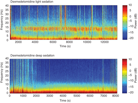
At clinically recommended doses used for light sedation, the EEG is characterized by spindles (intermittent bursts of 9- to 15-Hz oscillations) and slow–delta oscillations that resemble NREM stage II sleep (Figure 16.7).
FIGURE 16.7 Spectrogram of dexmedetomidine showing both spindles (9- to 15-Hz) and slow wave (0.1- to 1-Hz) oscillations at low doses and a predominance of slow-wave oscillations at high doses.
Two spectrograms visualizing frequency power over time during dexmedetomidine sedation: top shows light sedation, bottom shows deep sedation.
The slow oscillations seen with dexmedetomidine likely result from decreased excitatory inputs to the cortex (owing to the disinhibition of inhibitory circuits, as described in the previous section).
The spindles observed with dexmedetomidine, like sleep spindles and propofol-induced alpha oscillations, are thought to be generated by thalamocortical mechanisms. Of note, spindles are brief intermittent bursts of EEG activity, in contrast to propofol alpha oscillations, which are continuous.
These slow oscillations and spindles are smaller in amplitude than the slow and alpha oscillations observed with propofol general anesthesia, likely reflective of a lower level of circuit inhibition and thus lighter levels of sedation.
Molecular mechanism
Ketamine is an NMDA receptor antagonist that binds to receptors within the brain and spinal cord. Ketamine also interacts with opioid, monoaminergic, cholinergic, nicotinic, and muscarinic receptors.
Neural circuit mechanism
At low to moderate doses, ketamine primarily inhibits excitatory glutamatergic inputs to inhibitory interneurons, thereby disinhibiting downstream excitatory neurons. This can lead to increased cerebral
metabolism, as well as neuroexcitatory effects like hallucinations, dissociative states, and euphoria.At high doses, ketamine will also block the NMDA receptors on the excitatory glutamatergic neurons and lead to loss of consciousness.
The antinociceptive effect of ketamine is likely due to its actions on NMDA receptors in the dorsal root ganglia and non-NMDA mechanisms (eg, monoaminergic) in supraspinal regions.
EEG signatures
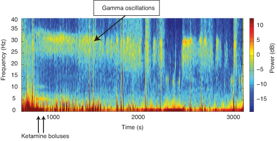
In patients receiving ketamine, the EEG is characterized by beta–gamma oscillations, ranging approximately between 20 and 30 Hz, in addition to the slow–delta oscillations characteristic of other anesthetics (Figure 16.8).
FIGURE 16.8 Spectrogram of ketamine showing notable 30-Hz gamma oscillations.
A frequency-time plot showing gamma oscillations and ketamine boluses. Color indicates power (dB).
For this reason, the numeric EEG-based index value associated with a ketamine anesthetic is often high, despite adequate anesthesia.
Nitrous oxide
Molecular mechanism
The mechanism of nitrous oxide is not completely understood, as it acts on many receptors. However, its anesthetic effects seem to be primarily due to antagonism of NMDA receptors.
EEG signatures
One common practice is to transition from an inhaled anesthetic (eg, sevoflurane, isoflurane) to high-concentration nitrous oxide (>50%) at the end of surgery to facilitate rapid emergence from anesthesia.
During this transition to nitrous oxide, the EEG is characterized by transient, prominent slow–delta oscillations (lasting 3 to 12 minutes), followed by high-frequency beta–gamma (20- to 35-Hz) oscillations that are more commonly associated with nitrous oxide alone (Figure 16.9).
FIGURE 16.9 Spectrogram of nitrous oxide during a transition from isoflurane as the anesthetic to nitrous oxide, as may occur clinically toward the end of surgery to facilitate emergence.The spectrogram of nitrous oxide profound slow–delta oscillations that transition to gamma oscillations.
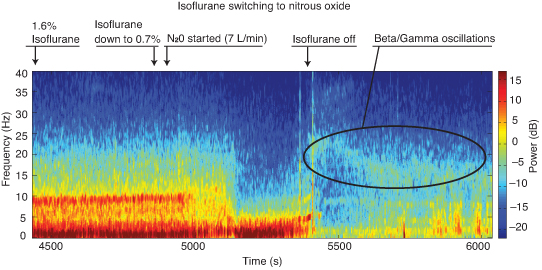
Spectrogram showing brain oscillations during anesthesia, labeled with drug changes: isoflurane (1.6% to 0.7% to off) and nitrous oxide. Color indicates power in decibels.
The beta–gamma oscillations observed under nitrous oxide are thought to be induced by a similar mechanism to ketamine, owing to the shared mechanism of NMDA antagonism.
The mechanism underlying the transient slow–delta oscillations is not fully understood.
Sevoflurane plus ketamine
Patients will often receive a combination of medications under anesthesia, such as an inhaled anesthetic like sevoflurane for unconsciousness and ketamine for analgesia.
When ketamine is added to sevoflurane general anesthesia, the alpha and delta oscillations typically observed with sevoflurane are reduced in power, whereas beta oscillations are increased in power. These observed changes may be due to ketamine causing an excitatory effect on neurons with lower intrinsic spiking frequencies to higher-frequency spiking.
Of note, the use of ketamine as an adjunct in a patient already receiving an inhaled anesthetic or propofol may lead to burst suppression.
Although not all combinations of anesthetics and their adjuncts have been studied at this time, understanding their mechanisms and the neural circuits involved can help provide insight into predicted or observed EEG patterns, as well as possible limitations with certain combinations of medications.