The body is very careful to prevent uncontrolled cell proliferation, as well as the replication of damaged DNA, so it has many safeguards in place. One of them is the ubiquitin–proteasome system. It is clear that proteins are the key to all cellular processes. The genes that code for them are the instructions or recipe to make the protein. Some proteins are extremely important, such as the cyclins and cyclin-dependent kinases that regulate the cell cycle. The cell cycle is turned on when certain proteins are made and their level in the cell rises, but then the cell cycle needs to be turned off; thus, these proteins need to be destroyed so they cannot be used to turn the cell cycle back on for proliferation without the usual checkpoints. Proteins also have time-limited function in signal transduction (kinases), differentiation, and DNA repair, and these proteins need to be turned on and off.
There are two main ways the body breaks down damaged proteins or proteins that are no longer needed, such as those just described. The first way is with lysosomes, which degrade extracellular proteins. The second way, with proteasomes, does not use lysosomes and is used to remove endogenous proteins the cell has made inside the cell. How does the proteasome know which proteins to degrade so that the protein can stop working (i.e., stop turning the cell cycle), and the components of the protein (e.g., amino acids) can be recycled? Ubiquitin, which means found everywhere, is a small regulatory protein that attaches to the proteins that need to be destroyed and recycled. This process is found in all our cells, and in fact, in all plants and animals (eukaryotic cells). It is a highly conserved process that allows the tight regulation of the concentration of specific proteins so that critical processes can be turned off, such as the cell cycle, and signal transduction. In addition, it is important in destroying proteins that are damaged or misfolded, such as proteins that have been made by mutated genes. It is also important in regulating gene expression. Ubiquitin tags are attached to the shortlived protein, and there are actually three different enzymes that help to tag the
protein (E1, E2, and E3). Once the tagging is complete and identified with its ubiquitin tag, the protein is escorted to the 26S proteasome, where it is degraded. The ubiquitin is released so it can go do the same job somewhere else in the cell.
There are many different proteasomes. The number of E3 molecules on the ubiquitin tag tells the protein which proteasome to go to. The 26S proteasome is one of the most important proteasomes. It is shaped like a barrel and is found both in the cell nucleus and in the cytoplasm. The barrel contains four stacked rings, called  or
 subunits, which surround a core. The tagged protein is recognized by the ends of the barrel (called regulatory caps) and pulled in. One of the subunits helps the protein move into the center core, the protein is unfolded, and the subunit then begins to degrade the protein. The protein is cut into peptides, by destroying the peptide bonds, and then further divided into the component amino acids that were used to make the protein in the first place. ATP is the cell's energy source that is used to degrade the protein. These amino acids are then released and reused (recycled) to make new proteins.
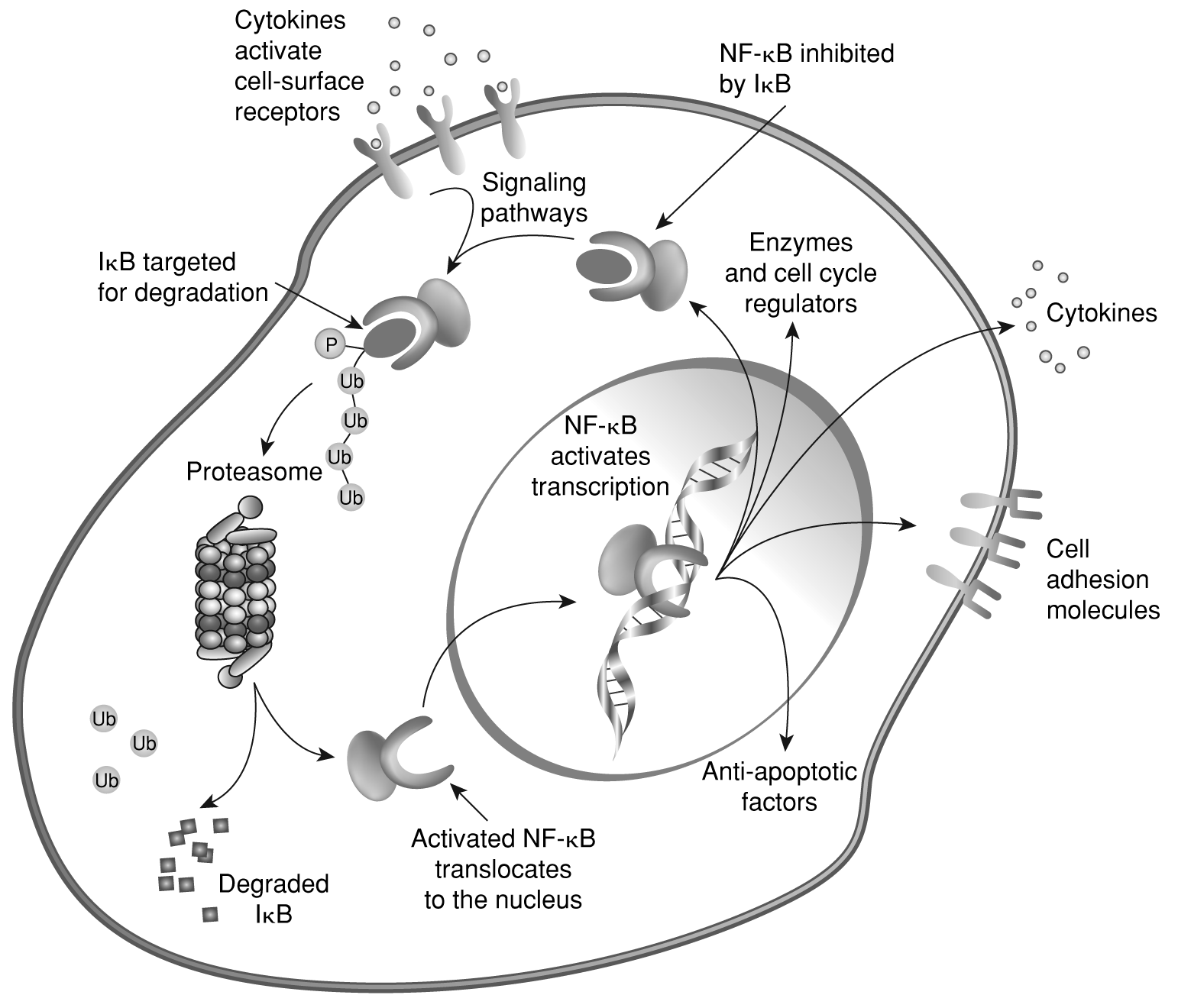
Cells depend on effective ubiquinated proteasome degradation of key proteins used in running the cell. When it is inhibited by a proteasome inhibitor such as bortezomib (Velcade), the cell goes crazy and dies. On a molecular level, it appears to cause increased levels of IkB, an inhibitor of nuclear factor-kappaB (NF-kB).149 Because it is not doing its job, misfolded proteins accumulate, ringing an alarm within the cell (unfolded protein response, which triggers endoplasmic reticulum stress). When the alarm is triggered, the cell interprets it as severe stress and initiates apoptosis. Interestingly, it was thought that green tea might enhance the effective cell killing started by bortezomib. However, the reverse was found, in that the green tea extract blocked proteasome inhibition by bortezomib; thus, patients taking bortezomib should not drink green tea or take green tea extracts.149 Figure 2-10 shows the ubiquitin–protease pathway and the interaction with NF-κB. The protein-degradation pathway is very tightly regulated so that cells can quickly turn on or off proteins necessary to keep the cell functioning optimally. In some cancers that have mutations in tumor suppressor proteins, such as BRCA1, which is involved in DNA repair, there is a mutation disabling ubiquitin from linking to the mutated tumor suppressor protein; thus, it is not inactivated and continues to allow division of cells with damaged DNA. In addition, there are oncogenes (e.g., double minute 2 [MDM2] and S-phase kinase protein [SKP2]) that send messages for ubiquitin to tag proteins, such as p53 and p27, that regulate the cell cycle and would otherwise stop the cell from dividing.150
HSPs are also in this group of potential targets for anticancer therapy.152 The body is very careful to have a rapid response team to help when the body is exposed to high temperatures or internal stress. Heat or stress causes the release of transcription factors that increase the expression of the HSPs (which is part of the heat shock response), mediated by heat shock factor (HSF). HSF is the
Figure 2-10. Ubiquitin-protease pathway and interaction with NF-B.
Lee Moffitt Cancer Center and Research Institute Inc.151
same molecule found in all living organisms, so it is a primitive protective mechanism for proteins. One has only to fry an egg to see the effects of heat on protein. HSPs play a role as molecular chaperones when they help fold proteins (they have to be folded correctly or they are unable to do their job), helping proteins cope with stress (e.g., inflammation, hypoxia), intracellular trafficking, and housekeeping (i.e., monitor protein health and help bring old proteins to the recycling bin or proteasome).152
Heat shock factor 1 (HSF1) is a transcription factor that upregulates (turns on the production) Hsp70 protein expression. Hsp70 and Hsp90 are HSPs that are highly expressed in cancer cells and are critical to the cancer cell's survival.153 Interestingly, Hsp70 not only protects the proteins within the cell, but it also helps the cell survive. It does this by (1) helping keep proteins folded correctly as they are being synthesized, (2) helping to bring proteins through the cell membrane in a partially folded state, (3) protecting them from thermal and oxidative stress, (4) interacting with E3 ubiquitin ligase to bring old proteins to the proteasome, and lastly, and most importantly, (5) directly inhibiting apoptosis. It does this by blocking procaspase-9 to Apaf-1/dATP/chytochrome c in the second step of the
cytochrome c pathway of apoptosis.154 Hsp90 is also a molecular chaperone that is highly expressed in most normal as well as cancer cells and is necessary for cell survival. Similar to Hsp70, Hsp90 is able to clamp onto proteins to help in protein folding, cell signaling, protein degradation, and protein protection from stress (e.g., hypoxia, free radicals, radiation, chemotherapy). Some of the proteins that Hsp90 chaperones are EGFR, ErbB2 (HER2), c-Met, BCR-Abl, RET, CDK4, FLT3, androgen receptors, B-Raf, NF-kB, C-Raf, p53, and HIF-1, which helps one appreciate how important this protein is as a target in cancer therapy.3 On one hand, Hsp90 protects the integrity of proteins by making sure that damaged or mismade proteins are brought to S26 proteasome for degradation by the ubiquitin– proteasome pathway. However, on the other hand, Hsp90 appears to protect damaged proteins, as elevated levels of Hsp90 allow the cell to accumulate the protein products of mutated genes as well; this may be an evolutionary advantage. However, in the context of cancer, this function underscores how important Hsp90 is as an anticancer target.
Hsp90 is necessary to keep the 26S proteasome functioning effectively, as it is responsible for the ATPase activity of the proteasome.155 However, even though it keeps the 26S proteasome in excellent functional shape, it is also able to help cancer protein kinases to survive in the cell cytoplasm (P13K-Akt). Not surprisingly then, Hsp is overexpressed in cancer cells. P13K and AKT are important proteins in cancer signal transduction, and if they are inhibited, the cell dies (undergoes apoptosis). Hsp90 stabilizes P13K and AKT proteins, so if Hsp90 can be blocked, it appears both P13K and AKT are also blocked, leading to cell death. Hsp90 is a good target for other reasons as well. Hsp90 protects or stabilizes mutated proteins such as the fusion oncogene BCR-Abl and p53, which is mutated in more than 50% of cancers.156 In addition, Hsp90 plays an important role in angiogenesis. It is necessary for VEGF and the endothelial cell to make and release nitric oxide, both of which are necessary for angiogenesis.157 Because of its wide-reaching effect on proteins, it influences tumor cell apoptosis (via Akt), TNF receptors, and NF-kB function, and through interaction with MMP2, it promotes invasiveness of the tumor so that it can metastasize.158
Clearly, Hsp70 and Hsp90 are attractive anticancer targets for three reasons.3 First, targeting Hsp90 will leave most normal proteins alone, as tumor cells have increased expression of Hsp90 and drugs targeted to Hsp90 should accumulate in the tumor cells. Second, because Hsp90 chaperones so many important cellular proteins, many of which are signaling proteins in malignant pathways, multiple targets can be attacked by just targeting Hsp90. Third, as we have seen, there are multiple signaling pathways to accomplish the same goal. Thus, by targeting multiple pathways mediated by Hsp90, the likelihood of the tumor cells escaping, as they would with a single targeted pathway, lessens. Conceptually, targeting both Hsp90 and the ubiquitin–proteasome pathway also makes excellent sense. This results in a pile of unfolded proteins that become toxic to tumor cells.159
Despite a strong argument for targeting Hsp90, little success has been found so far. More work has been done studying Hsp90 and developing agents that target this protein. A geldanamycin analogue, 17-AAG, is a Hsp90 inhibitor that promotes the degradation of oncoproteins HER2, mutant p53, c-Raf, BCR-Abl.3 The dose-limiting side effects are anemia, thrombocytopenia, dehydration, hyperglycemia, and GI effects. Other common toxicities are fatigue, anorexia, diarrhea, nausea, and vomiting.3 Other agents being studied are tanespimycin (KOS-953), which is in phase III (for GIST, myeloma) and phase II (for pancreatic and renal cancer) testing, as well as retaspimycin (IPI-504), which is being studied in phase II trials (for GIST, NSCLC, prostate cancer). Alternatively, another strategy to attack the HSPs is via epigenetic routes, with HDAC inhibitors.3
Drug: Bortezomib (Velcade)
Indications: Bortezomib is FDA approved for the treatment of patients with:
- Multiple myeloma
- Mantle cell lymphoma who have received at least one prior therapy
Class: Proteasome inhibitor
Mechanism of Action: A reversible inhibitor of the 26S proteasome; inhibits the breakdown of ubiquinated intracellular proteins and disrupts the ubiquitin– proteasome pathway. This pathway normally regulates the intracellular concentration of specific proteins, thus controlling homeostasis. Cancer cells depend on the proteins that are available from this process to turn on the cell cycle and to make the apparatus of mitosis. When the ubiquitin–proteasome pathway is disrupted, the proteins are not available, and multiple signaling pathways within the cell are disrupted, encouraging the cell to undergo apoptosis. Cell cycle movement (cell division) stops. Cells are unable to migrate, and sensitivity to chemotherapy is increased. In addition, the drug appears to downregulate the NF-κB pathway, which is necessary for cell growth, avoidance of apoptosis, and adhesion. This downregulation may restore chemosensitivity. In multiple myeloma, it interferes with cellular adhesion molecules so that tumor cells cannot bind to the bone marrow.
Pharmacokinetics: After IV administration, the drug is rapidly cleared from the plasma, with a mean elimination half-life range of 40–193 hours after multiple dosing. Drug undergoes oxidative metabolism via cytochrome P450 enzymes 3A4, 2D6, 2C19, 2C9, and 1A2. Drug is deboronated into two metabolites that are then hydroxylated into several metabolites. The elimination path is unknown.
Dosage and Administration: 1.3 mg/m2 administered as a 3- to 5-second bolus IV injection. Specific conditions as follows:
Multiple myeloma, previously untreated: Administered in combination with oral melphalan (9 mg/m2) and prednisone (60 mg/m2) given PO on days 1–4 of a 6-week cycle.
- Cycles 1–4, bortezomib is administered once weekly (days 1, 4, 8, 11, 22, 25, 29, and 32).
- Cycles 5–9, bortezomib is administered once weekly (days 1, 8, 22, and 29).
- At least 72 hours should elapse between consecutive doses of bortezomib.
- Dose modifications: For prolonged grade 4 neutropenia or thrombocytopenia with bleeding in the previous cycle, consider reduction of melphalan dose by 25% in next cycle. If platelet count is
 30,000/mm3 or ANC 750/mm3 on a bortezomib dosing day other than day 1, bortezomib dose should be held.
- If bortezomib doses in consecutive cycles are held due to toxicity, bortezomib dose should be reduced by one dose level (from 1.3 mg/m2 to 1 mg/m2, or from 1 mg/m2 to 0.7 mg/m2).
- Grade 3–4 nonhematologic toxicities: Hold bortezomib until symptoms have resolved to grade 1 or baseline. Bortezomib may then be reinitiated with one dose level reduction (from 1.3 mg/m2 to 1 mg/m2, or from 1 mg/m2 to 0.7 mg/m2). For bortezomib-related neuropathic pain and/or peripheral neuropathy, hold or modify bortezomib.
Relapsed multiple myeloma or mantle cell lymphoma: Bortezomib is given twice weekly for 2 weeks (days 1, 4, 8, and 11) followed by a 10-day rest period (days 12–21).
- For extended treatment of more than 8 cycles, bortezomib may be given on the standard or maintenance schedule of once weekly for 4 weeks (days 1, 8, 15, and 22) followed by a 13-day rest period (days 24–35).
- At least 72 hours should elapse between consecutive doses of bortezomib.
- Dose modifications: Hold bortezomib for any grade 4 nonhematologic or grade 4 hematologic toxicity except for peripheral neuropathy, which is discussed separately. Once the toxicity symptoms have resolved, resume bortezomib at a 25% dose reduction (e.g., 1.3 mg/m2 to 1.0 mg/m2, or 1 mg/m2 to 0.7 mg/m2).
Peripheral neuropathy dose modifications:
- Grade 1 (paresthesias, weakness +/– loss of reflexes) without pain or loss of function: No action is taken.
- Grade 1 with pain or grade 2 (interfering with function but not with ADLs: Reduce bortezomib to 1 mg/m2.
- Grade 2 with pain or grade 3 (interfering with ADLs): Hold bortezomib until toxicity resolves. When symptoms resolve, reinitiate with a reduced dose of bortezomib (0.7 mg/m2), and change schedule to once a week.
- Grade 4 (sensory neuropathy that is disabling or motor neuropathy that is life-threatening or leads to paralysis): Discontinue bortezomib.
Available as: One single-use 10-mL vial contains 3.5 mg of bortezomib. Dose must be individualized to prevent overdose.
Reconstitution and Preparation: Store unopened vials at room temperature, 25°C (77°F), and protect from light. Reconstitute each vial with 3.5 mL 0.9% sodium chloride injection USP, forming a colorless and clear solution (stable for 8 hours at controlled room temperature). Final concentration is 1 mg/mL of bortezomib. Draw up prescribed amount (stable in syringe for 8 hours).
Drug Interactions:
- Potent CYP3A4 inhibitors (e.g., ketoconazole, ritonavir): Increase bortezomib exposure; monitor closely for toxicity or reduce dose of bortezomib.
- Patients receiving other drugs that are either inhibitors or inducers of CYP3A4 should be monitored closely for toxicity or reduced efficacy.
- In clinical trials, the addition of dexamethasone 20 mg PO given the day of and the day after bortezomib resulted in increased response rate in patients with stable disease or progressive disease on bortezomib alone.
- Green tea: Polyphenols in green tea may decrease the drug's ability to induce cancer cell death and can negate the drug's efficacy.
Contraindications: Patients with hypersensitivity to bortezomib, boron, or mannitol
Warnings:
- Women of childbearing age should use effective contraception to avoid becoming pregnant while being treated with bortezomib, as the drug decreases the chances of successful delivery of the fetus.
- Women should be advised against breastfeeding or becoming pregnant while being treated with bortezomib.
- Peripheral neuropathy is primarily sensory but may be severe. Patients should have a neurologic assessment prior to each drug dose, eliciting history (symptoms such as pain, burning, paresthesias, dysesthesias, weakness); patients should be asked to pick up a coin and squeeze the nurse's hand; and the nurse should observe patient walking into the exam room to see if there is any gait disturbance or if patient is holding onto the wall. If patient now wears sneakers, it may be an indication of numbness in the toes, but patient needs to validate this observation. Discuss your findings with physician or midlevel practitioner to determine dose modification or discontinuance depending upon severity. Patients report significant improvement in peripheral neuropathy grade after drug dose decrease or drug discontinuation, so peripheral neuropathy is largely reversible. Patients with preexisting severe neuropathy should be treated with bortezomib only after careful risk–benefit assessment.
- Hypotension can occur (incidence 13%). Caution should be used when treating patients receiving antihypertensives, those with a history of syncope, and those who are dehydrated. Teach patients to drink adequate fluid throughout the day (1–2 liters with one glass every hour while awake). If the patient has nausea and vomiting, or diarrhea, teach patient self-administration of antiemetics or antidiarrheal medication and to call if these measures are ineffective in 24 hours.
- Patients with risk factors for or existing heart disease should be closely monitored. There are rare reports of decreased LVEF, CHF, and QTc prolongation.
- Acute diffuse infiltrative pulmonary disease has been reported, as has alveolitis, interstitial pneumonia, lung infiltration, and ARDS. Rare reports of pulmonary hypertension have been made. If the patient has worsening of any cardiac function, a prompt cardiovascular workup should occur.
- Nausea, diarrhea, constipation, and vomiting have occurred and may require use of antiemetic and antidiarrheal medications or fluid replacement.
- Thrombocytopenia or neutropenia can occur; CBC should be regularly monitored prior to each dose and throughout treatment.
- TLS, reversible posterior leukoencephalopathy syndrome (RPLS), and acute hepatic failure have been reported. For TLS, patients with high tumor burden who are beginning therapy should receive adequate TLS prophylaxis. For RPLS, ensure patients have well-controlled hypertension. If patient has change in mental status, confusion, lethargy, seizure, blindness, or other neurologic symptoms, stop bortezomib and evaluate the patient thoroughly. MRI will be diagnostic for RPLS. Manage with control of BP. See Chapter 3 for more on antiangiogenic agents. For acute hepatic failure, assess LFTs baseline and periodically during treatment. This condition appears to be reversible with drug discontinuance.
- Patients with diabetes may require close monitoring of blood glucose and adjustment of antidiabetic medication.
note:Adverse Reactions (bold indicates most common, occurring in > 25% of patients):
Cardiovascular: Hypotension, orthostatic hypotension, peripheral edema, hypertension
Pulmonary: Dyspnea, upper respiratory infection (URI), cough, pneumonia, bronchitis
CNS/neurologic:Peripheral neuropathy, psychiatric disorders, neuralgia, headache, insomnia, dizziness, blurred vision
Dermatologic:Rash,pruritus
Metabolic:Increased hyperglycemia in patients with diabetes mellitus
GI:Diarrhea, moderate potential for nausea and vomiting, constipation, anorexia, dysgeusia, dehydration, abdominal pain
Hematologic:Thrombocytopenia with nadir day 11 and recovery by day 21; neutropenia, leucopenia, anemia
Musculoskeletal: Back pain, pain in extremity, arthralgias, myalgias
General:Pyrexia, asthenia, rigors
Nursing Discussion:
Peripheral neuropathy: Of patients in clinical trials, 80% had a preexisting baseline peripheral neuropathy (PN); 37% of patients had new onset or aggravation of existing PN; and 14% developed grade 3 PN overall, with 5% occurring in patients without baseline PN symptoms. There were no patients with grade 4 PN. PN improved/resolved in 51% of patients who underwent a dose adjustment for grade 2 PN and in 73% of patients who discontinued the drug. Teach patient to report new onset or worsening of PN symptoms (paresthesias, dysesthesias), any changes in sensory function (temperature sensation, knowing where body parts are in relation to the whole, etc.), functional ability (especially senses of smell and taste), and ability to carry out ADLs. Assess severity of symptom(s) if they arise and potential for injury. If moderate, consult protocol and discuss medical intervention with physician. Teach patient measures to minimize symptoms and ensure safety. Discuss coadministered medication profile with physician, as concomitant administration of the following drugs increases risk of PN: amiodarone, antivirals, isoniazid, nitrofurantoin, or statins. Discuss adding agents that may decrease development or worsening of PN, such as glutamine. See Dosage and Administration.
note:Bone marrow depression with thrombocytopenia and neutropenia: Thrombocytopenia occurred in 43% of patients (27% grade 3 and 3% grade 4, with nadir day 11 and recovery by day 21) and neutropenia in 24% of patients (13% grade 3 and 3% grade 4; incidence higher in patients with multiple myeloma than mantle cell lymphoma). Febrile neutropenia occurred in < 1% of patients. Anemia incidence was 32%. Fever is common and higher in patients with multiple myeloma (37%) compared with mantle cell (19%). Assess baseline leukocyte, platelet, and Hgb/HCT, and monitor before each treatment, during therapy, and more often as needed. Hold drug for ANC < 1000/mm3, and dose reduce 25% (see Dosage and Administration). Assess risk for infection and integrity of skin and mucous membranes, pulmonary status, and ability to clear secretions, as well as history of past infections, baseline and prior to each treatment. Teach patient to self-assess for signs/symptoms of infection and to call provider immediately or come to the emergency department if temperature > 100.5°F, shaking chills, rash, productive cough, burning on urination, or any signs/symptoms of infection or bleeding. Teach self-care strategies to minimize risk of infection and bleeding, including avoidance of OTC aspirin-containing medications.
Increased risk of herpes zoster: Risk of reactivation of herpes zoster in both newly diagnosed and relapsed multiple myeloma patients was 13%.160 The incidence of herpes simplex was 2–8% in the patients receiving bortezomib compared to 1–5% in the control group. When patients received prophylactic antiviral
therapy, previously untreated multiple myeloma patients had a decreased incidence (3%) compared to those who did not (17%). Rare cases of herpes meningoencephalitis and ophthalmic herpes have been reported postmarketing. Thus it is recommended that physicians consider prophylactic antiviral therapy.160 161 For example, in some institutions, acyclovir 400 mg PO twice daily is used if the patient's renal function is normal.
Physicians should consider using antiviral prophylaxis in subjects being treated with Velcade. In the randomized studies in previously untreated and relapsed multiple myeloma, herpes zoster reactivation was more common in subjects treated with Velcade (13%) than in the control groups (4–5%). Herpes simplex was seen in 2–8% of subjects treated with Velcade and 1–5% in the control groups. In the previously untreated multiple myeloma study, herpes zoster virus reactivation in the Velcade, melphalan, and prednisone arm was less common in subjects receiving prophylactic antiviral therapy (3%) than in subjects who did not receive prophylactic antiviral therapy (17%). In the postmarketing experience, rare cases of herpes meningoencephalitis and ophthalmic herpes have been reported.
Nausea, vomiting, decreased appetite, diarrhea, constipation, and dehydration: Patients in clinical studies developed these symptoms with the following incidence: nausea (64%), diarrhea (51%), decreased appetite (43%), constipation (43%), vomiting (36%), dehydration (18%). Diarrhea, nausea, and vomiting occur 6–24 hours after infusion. Nausea is more common in patients with multiple myeloma. Diarrhea and constipation may occur during cycles 1 and 2, and then disappear. Patients with diabetes require close monitoring of their blood glucose and may require adjustments in their antidiabetic medication. Assess baseline weight and nutritional status, and monitor prior to each treatment. Assess glucose and electrolytes prior to each treatment to weekly, especially serum sodium and potassium; replete as necessary, and teach diet high in sodium and potassium. Teach patient that serum electrolytes may be decreased and to assess for signs/symptoms of hyponatremia (confusion, weakness, seizures), hypokalemia (muscle weakness, confusion, irregular heartbeats), hypercalcemia (constipation, thirst, confusion, muscle cramps, sleepiness), and hypomagnesemia (muscle cramps, headache, weakness). Teach patient that side effects may occur, self-care strategies, and to report them if symptoms do not resolve. Use aggressive antiemetics to prevent nausea and vomiting: serotonin antagonist IV prior to bortezomib dose and for 36 hours after each drug dose, if needed. If diarrhea develops after first treatment, teach patient to take loperamide prior to next dose of bortezomib and after every loose stool for 36 hours (not to exceed 8 tablets a day), as well as to use the bananas, rice, applesauce, toast diet. If patient develops constipation, teach preventive self-care (stool softener, flax-seed oil, Milk of Magnesia, prunes or prune juice every a.m. to ensure bowel movement at least every other day). Teach patient to drink 2 quarts of fluid daily, drinking 1 glass an hour while awake to prevent dehydration. Teach patient to report dizziness, lightheadedness, or
fainting spells, and to avoid operating heavy machinery or driving a car if these occur. Develop symptom management plan with physician. Make referral to dietitian as appropriate. Teach diabetic patients to monitor their blood glucose closely, and discuss abnormalities and changes in their antidiabetic dose with their nurse practitioner, physician's assistant, or physician.