▶Also known as lobular capillary hemangioma.
▶Common in children and young adults.
▶Acquired vascular lesion of skin or mucous membranes.
▶Cause unknown but appears to represent reactive neovascularization.
▶Solitary red papule or papulonodule, rarely larger than 1 cm.
▶May be pedunculated or exophytic (Figure 60.1).
▶Surface commonly bleeds or becomes erosive or crusted (Figure 60.2).
▶Base of lesion may be surrounded by collarette of scale.
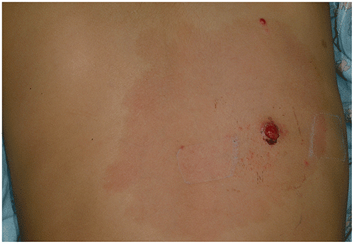
▶May develop on surface of port-wine stain (Figure 60.3).
▶Occasionally multiple.
▶Common locations include hand, finger, face, and oral mucosa.
Figure 60.1. Pyogenic Granuloma. An Eroded, Exophytic Vascular Papule with Peripheral Collarette of Scale, on the Finger of a 14-Year-Old.
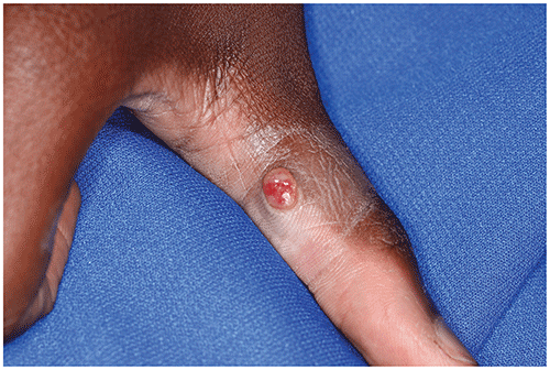
Figure 60.2. Pyogenic Granuloma. This Multi-Lobulated, Vascular Papule Was Prone to Recurrent Bleeding and Crusting (as Noted at Superior Portion).
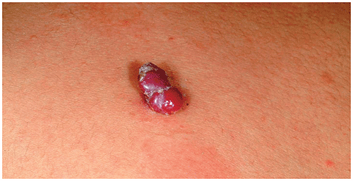
Figure 60.3. Pyogenic Granuloma Overlying Port-Wine Stain. Note 2 Vascular Papules, One at the Superior Pole and the Other (Eroded) More Centrally Located.
Look-alikes
| Disorder | Differentiating Features |
|---|
| Infantile hemangioma | |
| Juvenile xanthogranuloma | Early lesion may appear vascular, but eventually yellow-orange hue becomes apparent. Usually sessile (broad-based) rather than pedunculated. Rarely becomes erosive or crusted on surface.
|
| Spitz nevus | Slowly growing papule. Sessile (broad-based) rather than pedunculated. Rarely becomes erosive or crusted on surface. Diascopy (pressure with glass slide) may reveal brown pigment. Dermoscopy (examination with a dermatoscope) may reveal characteristic pigment patterns.
|
| Spider angioma | May simulate early (small) pyogenic granuloma. Central papule remains < 3 mm. Does not become erosive on surface. Peripheral telangiectatic vessels present.
|
▶Pyogenic granuloma is usually diagnosed based on the classic clinical features.
▶Histologic evaluation is confirmatory after excision.
▶Shave excision followed by electrocautery of the base.
▶Small lesions may be amenable to pulsed dye laser therapy, topical timolol gel.
▶Very small lesions with eroded surface may respond to chemical cauterization with silver nitrate.
▶Full-thickness excision occasionally indicated for larger lesions.
▶The prognosis for an uncomplicated pyogenic granuloma is excellent.
▶Recurrence is rare but may occur after excision.
▶Patients with multiple, clustered (agminated) lesions are more prone to recurrence.
▶Consider referral when the
■Diagnosis is in question.
■Patient or parent desires removal.
■Lesion is erosive or bleeding.