▶Pigmented band of the nail unit; also known as longitudinal melanonychia.
▶May present at any age, including in children.
▶Common in individuals with skin of color, especially Black individuals (present in up to 77%, with nearly 100% developing the finding by 50 years of age; often involves multiple digits); seen in up to 20% of Japanese individuals and approximately 1% of white individuals.
▶Underlying disease may represent melanocytic activation or melanocytic hyperplasia.
▶May represent either a benign (ie, lentigo or benign nevus) or malignant (ie, melanoma) process, despite similar clinical presentations in both settings.
▶Approximately 5% to 10% of cases in adults result from a subungual melanoma; subungual melanoma as a cause for melanonychia is rare in children.
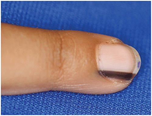
▶Longitudinal, pigmented (tan, brown, dark brown, or black) streak of the nail plate (Figures 75.1 and 75.2).
▶May or may not have associated nail deformity.
▶Hutchinson sign (melanin pigmentation extends to skin and soft tissue proximal to the nail plate) is traditionally considered pathognomonic for subungual melanoma. However, noting some periungual pigmentation is not necessarily a positive Hutchinson sign; pseudo-Hutchinson sign (reflection of pigmentation through the cuticle/nail fold) is reported more frequently in pediatric patients.
▶Other concerning features for malignancy include sudden or rapid change in the width of the pigmented band and irregular or blurred borders (see
When to Worry or Refer
section).
Figure 75.1. Solitary, Well-Demarcated, Light Tan Longitudinal Band of the Thumb in an 8-Year-Old.
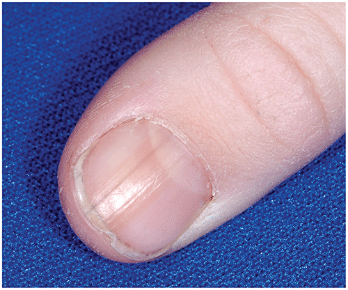
Figure 75.2. Very Dark-Brown Linear Streak on the Index Finger of a 5-Year-Old. This Lesion Continued to Change and Was Eventually Biopsied, Revealing a Benign Nevus in the Nail Matrix Area.
Look-alikes
| Disorder | Differentiating Features |
|---|
| Physiologic melanonychia | Commonly caused by chronic local nail trauma (ie, nail biting and cuticle trauma during a pedicure). Other causes include pregnancy, certain drugs, some systemic diseases. May affect multiple nails, especially in individuals with skin of color.
|
| Onychomycosis | Dermatophyte fungal infection of the nail plate. May rarely be associated with partial or complete melanonychia of one or more nails. Typically associated with a thickened nail plate that may separate from the nail bed, as well as yellow or white discoloration of nail plate. Treatment for fungal infection results in reversal of nail discoloration.
|
| Addison disease | Adrenal insufficiency may darken skin, mucosae, and nails. Longitudinal melanonychia can be the first (and occasionally only) sign of Addison disease. Nail pigmentation disappears after treatment of the primary disease.
|
| Laugier-Hunziker syndrome | Idiopathic macular hyperpigmentation on mucosal lips (<5-mm macules typically), oral cavity, and nails. Benign disease with no systemic involvement.
|
| Peutz-Jeghers syndrome | Combination of pigmentary macules (lips, around and inside mouth, perianal, hands, and feet) with predisposition toward intestinal hamartomatous polyposis. Variation of the SKT11 gene. Melanonychia of one or multiple nails can rarely be seen. Must evaluate for associated gastrointestinal polyps and other potential malignancies (with long-term surveillance).
|
▶The diagnosis is typically made clinically based on the typical appearance of a pigmented longitudinal streak of the nail plate.
▶The presence of multiple bands, as occasionally noted in patients with skin of color, is reassuring and typically lessens the likelihood of a malignant cause.
▶No treatment is necessary for benign melanonychia striata.
▶Avoid trauma, as feasible.
▶Rapid changes or sudden darkening in color must be assessed for possible malignant transformation.
▶When there is a clinical concern for possible malignancy, biopsy of the nail matrix for histologic evaluation is indicated.
▶Higher-grade atypical melanocytic lesions and melanoma are treated with complete surgical excision.
▶Melanonychia striata has an excellent prognosis when caused by a benign nail matrix lesion such as lentigo or benign nevus.
▶When subungual melanoma is present, the prognosis depends on a variety of histologic features as well as depth of the primary lesion.
▶Refer patients who have melanonychia striata with atypical features or a family history of melanoma to a dermatologist for evaluation and follow-up; additional evaluations may include dermoscopy and/or nail matrix biopsy.
▶Atypical features in melanonychia striata may include older age (older adults are at far greater risk for subungual melanoma than children are), rapid evolution, greater (>3 mm) width of the band, blurred borders, irregular pigmentation, extension of pigment onto adjacent skin (Hutchinson sign), having a mass under the nail plate (with lifting of the plate), bleeding or ulceration, and pain (if there is extension to bone).