▶Rare inflammatory vasculopathy primarily involving the skin and muscle with potential for multisystem compromise.
▶A small subset of patients has no evidence of muscle disease; termed dermatomyositis sine myositis (or amyopathic dermatomyositis).
▶Bimodal age peaks: childhood (5–10 years) and adulthood (45–55 years).
▶Incidence of approximately 2 to 4 cases per 1 million children per year.
▶Increased ratio of female to male patients in children and adults.
▶Overlap with scleroderma, juvenile dermatomyositis (JDM) or other connective tissue disease may occur in a minority of patients.
▶Although dermatomyositis in adult patients may be a marker for occult malignancy, this association is not seen in children.
▶Etiology and pathogenesis of JDM are poorly understood; believed to be autoimmune in nature, and patients may have familial, genetic predisposition.
Cutaneous
▶JDM has pathognomonic skin changes that may vary greatly in severity; characteristic inflammatory and telangiectatic skin findings are seen in the vast majority of children who are affected.
▶Pink to violet discoloration of eyelids (heliotrope) and cheeks in malar distribution with or without associated edema of affected skin (Figures 118.1 and 118.2).
▶Erythema may be present on the extensor aspects of the extremities (especially over elbows and knees), neck, over shoulders, and hairline.
▶A malar facial rash, similar to that seen in systemic lupus erythematosus, may occur.
▶Photosensitivity is common, with relative sparing of sun-protected sites.
▶Gottron sign or papules: pink to red telangiectatic macules (Gottron sign) or flat-topped lichenoid papules (Gottron papules), most often located over the proximal interphalangeal and metacarpophalangeal joints; less often involve the distal interphalangeal joints (Figures 118.3, 118.4, 118.5, and 118.6). These lesions may appear hypopigmented in darker skin tones.
■May be scaly.
■Occasionally appear symmetrically over the extensor aspects of the extremities (elbows, knees) and may resemble lesions of psoriasis (Figure 118.7).
▶Dilated capillaries (telangiectasias) of the proximal nail folds (may need magnification with ophthalmoscopy or dermatoscopy to visualize) (Figure 118.8). May also see areas of capillary dropout within these areas of capillary dilatation. These periungual changes appear to correlate with skin disease activity.
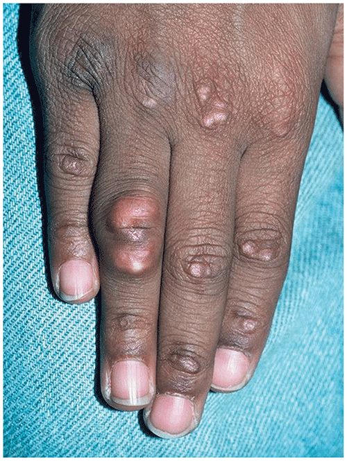
▶Calcinosis cutis occurs in 30% to 50% of children who are affected. Variably sized calcium deposits can present as firm skin-colored to white or yellow papules or nodules, usually located over the joints of the elbows or knees and the buttocks, but may occur anywhere (Figure 118.9).
■More common in pediatric than in adult patients.
■Usually a later finding; rarely seen at time of initial presentation.
■Occasionally become secondarily infected.
■Can become disabling if extensive.
■Severity of calcinosis seems to correlate with severity of inflammation and overall disease severity.
■Seen less frequently with aggressive and early therapy.
▶Widespread edema of the skin may be seen in more severe cases.
▶Poikilodermatous changes (ie, atrophy, telangiectasias, and hypopigmentation and hyperpigmentation within same region of skin) may be seen in chronic disease; often, there is a distinct violaceous discoloration of the skin.
▶Localized or widespread ulcerations may occur; extensive ulcerations believed to be associated with poor prognosis.
▶Inflammation of the scalp with associated scarring or non-scarring alopecia seen occasionally in children with JDM; more often seen in adults.
▶Lipodystrophy may occasionally be seen in association with panniculitis (inflammation of subcutaneous fat); may be generalized or partial.
▶Acanthosis nigricans (velvety hyperpigmentation of the neck, axillae) is occasionally seen in patients with JDM, especially those with lipodystrophy.
Figure 118.1. Heliotrope Rash and Telangiectatic Erythema of the Cheeks in a School-Aged Child with Juvenile Dermatomyositis.
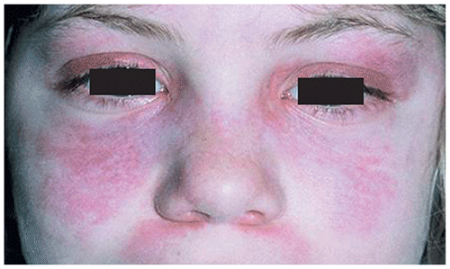
Figure 118.2. More Pronounced Erythematous to Violaceous Patches in Juvenile Dermatomyositis on the Face of a Child with a Darker Skin Tone Than the Patient Shown in Figure 118.1.
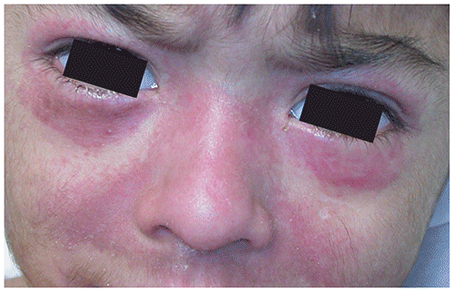
Figure 118.3. Typical Gottron Papules (Erythematous to Violaceous Flat-Topped Papules) Overlying the Knuckles in This 3-Year-Old with Juvenile Dermatomyositis. Note Also the Presence of Dilated Nail Fold Capillaries (See Figure 118.8).
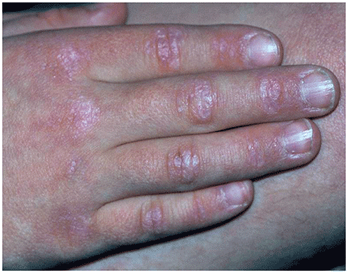
Figure 118.4. Gottron Papules Overlying Knuckles in a Child with Juvenile Dermatomyositis.
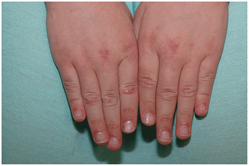
Figure 118.5. Juvenile Dermatomyositis. Gottron Papules on Bilateral Hands and Knees.
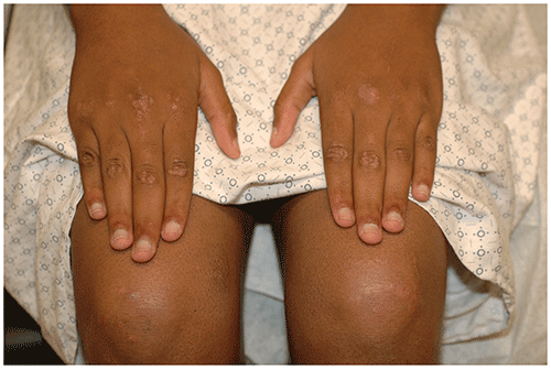
Figure 118.6. Numerous Gottron Papules in a 2-Year-Old Who Has Juvenile Dermatomyositis.
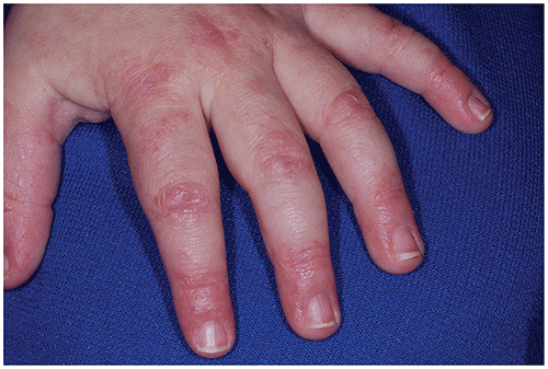
Figure 118.7. Erythematous Xerotic Papules on the Elbows in a Patient with Juvenile Dermatomyositis.
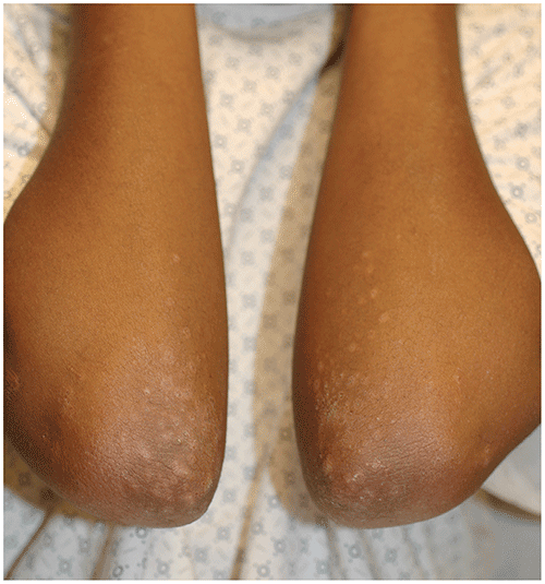
Figure 118.8. Dilated Capillaries of the Nail Folds (Arrows) in a Child with Juvenile Dermatomyositis.
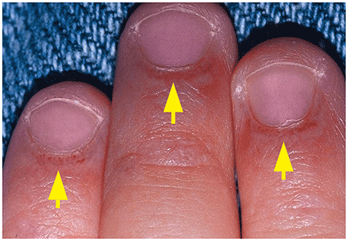
Figure 118.9. Calcinosis Cutis of the Fourth Finger as Well as Gottron Papules on the Knuckles in This Patient with a Long History of Juvenile Dermatomyositis.
Systemic
▶Symmetric proximal muscle weakness may precede, accompany, or follow skin changes.
■Usually involves the anterior neck flexors, the hip and shoulder girdles, and core musculature.
■May present with difficulty climbing stairs, raising arms to brush hair, or rising from lying to sitting and sitting to standing positions (also known as Gower sign).
■May or may not have associated muscle pain or tenderness to palpation.
▶May involve other striated muscle and result in symptoms of dysphagia, dysphonia, choking, or nasal speech.
▶In severe disease, can progress to involve respiratory muscles and lead to restrictive or interstitial lung disease.
▶Vasculopathy can occasionally lead to myocarditis, pericarditis, mucosal ulcerations of the gastrointestinal tract, or microscopic hematuria.
▶Fatigue and loss of energy are reported in most patients at presentation.
▶A nondestructive arthritis may occur in up to one-half of children who are affected.
▶Kidney involvement is more common in adults, and presence should prompt evaluation for lupus.
Look-alikes
| Disorder | Differentiating Features |
|---|
| Psoriasis | Psoriatic lesions of knees and elbows may resemble those of juvenile dermatomyositis (JDM) but usually contain thicker, micaceous (silvery white) scale. May have associated nail changes (eg, pitting, onycholysis). No dilated capillaries of nail folds. No calcinosis cutis. Facial involvement less common (but more common in pediatric psoriasis compared with that in adults). Histologic findings distinctive. Usually improves (rather than being exacerbated) with sun exposure.
|
| Allergic contact dermatitis | May have more marked edema of eyelids and affected skin. More acute onset than JDM. Severe pruritus usually present.
|
| Systemic lupus erythematosus | Usually less eyelid involvement. Distinct systemic manifestations. Photosensitivity a prominent feature, often with butterfly facial erythema (malar rash), with nasolabial sparing. Erythema of the dorsal fingers usually spares the areas over joints. Serological studies may help distinguish the 2 disorders.
|
| Scleroderma or CREST syndrome (calcinosis, Raynaud phenomenon, esophageal involvement, sclerodactyly, telangiectasia) | May have similar telangiectatic changes around the nails as in JDM. May have symptoms of dysphagia in both conditions. May also have calcinosis cutis in CREST syndrome. Sclerodactyly (thickening and tightness of the fingers and toes) or generalized induration not typically seen in JDM. Distinctive histologic changes at skin biopsy. JDM and scleroderma can present together as an overlap syndrome in children.
|
| Atopic dermatitis | Often with earlier onset (infancy or toddler years). Usually associated with more severe pruritus. Predilection for neck and flexural aspects of extremities (extensor surfaces in infants).
|
| Cutaneous T-cell lymphoma | Rare in children. Hypopigmented form most common in pediatric patients. Poikilodermatous form seen mainly in adults. Characteristic histologic features seen at skin biopsy.
|
| Postinfectious myopathy/myositis | |
| Collagen vascular disease–associated myositis or myopathy | May or may not have associated dermatologic alterations. Systemic lupus erythematosus–associated myositis generally does not have significant elevation of muscle enzymes. May have other systemic alterations not typically seen in JDM.
|
▶There is no single diagnostic test for JDM.
▶The diagnosis is suggested clinically by the combined findings of pathognomonic skin changes and associated symmetric proximal muscle weakness.
▶Supportive diagnostic evidence includes the following:
■Elevated skeletal muscle enzymes.
– Creatine phosphokinase.
– Aldolase.
– Aspartate aminotransferase, alanine aminotransferase, and lactate dehydrogenase; although generally characterized as liver enzymes, these may be elevated in dermatomyositis because they are released from damaged muscle tissue.
– Elevated level of inflammatory markers (ie, erythrocyte sedimentation rate, C-reactive protein level) may be present, but does not necessarily correlate with disease activity and is nonspecific.
■Characteristic histologic changes at skin biopsy (epidermal atrophy, interface dermatitis, mucin deposition).
■Characteristic histologic findings from muscle biopsy (usually deltoid or quadriceps). Although this procedure has been largely replaced by magnetic resonance imaging, which reveals increased signal intensity on fat-suppressed T2-weighted images, it should be considered for patients in whom the clinical presentation is not classic or pathognomonic and may provide prognostic information; magnetic resonance imaging may be helpful in following the clinical course of muscle involvement.
■Characteristic electromyographic findings.
■Some myositis-specific antibodies may be present and may help in elucidating prognosis.
▶Most patients are treated by pediatric rheumatologists; pediatric dermatologists are often involved at diagnosis, when patients present with cutaneous signs and symptoms.
▶Although muscle disease is usually quite responsive to therapy, cutaneous disease may be very resistant to multiple treatment modalities.
▶Persistent cutaneous disease may be associated with long-term cardiovascular risk.
▶Mainstays of treatment include the following:
■Photoprotection.
– Daily use of broad-spectrum sunscreen with sun protection factor 30 or higher.
– Use of sun protective clothing (including wide-brimmed hats).
– Avoidance of prolonged sun exposure.
■Topical steroids or topical calcineurin inhibitors (eg, pimecrolimus, tacrolimus) may be helpful for any associated pruritus and erythema but rarely modify the course of cutaneous disease.
■Systemic corticosteroids (oral or pulsed intravenous [IV]); specialists who treat JDM are increasingly using high-dose pulsed IV steroids.
■Immunosuppressive therapy and steroid-sparing agents.
– Methotrexate (oral or subcutaneous)
– Hydroxychloroquine (low dose), often used for inflammatory skin disease (although effectiveness is controversial)
– IV immunoglobulin
– Cyclosporine
– Mycophenolate mofetil
– Tacrolimus
– Pulsed cyclophosphamide
– Rituximab
– Tumor necrosis factor α antagonists including infliximab, adalimumab, and etanercept (although some patients appear to worsen with these therapies)
– Abatacept (recombinant DNA–generated fusion protein)
– Janus kinase inhibitors
■Autologous stem cell transplant (in some severe cases).
■Physical therapy.
▶Arthritis, gastrointestinal tract vasculopathy with ulceration or hemorrhage, malabsorption, and interstitial lung disease may occur and require specific evaluation as indicated.
▶Calcinosis cutis is very difficult to treat; reported therapies include increasing systemic immunosuppression, bisphosphonates, sodium thiosulfate, surgery.
▶The prognosis is variable but seems quite favorable for most children treated aggressively and early within the disease course. Many will become disease free after 2 to 4 years and remain so after therapy.
▶Control of skin disease and muscle disease does not correlate well; muscle disease is usually quite responsive to corticosteroid therapy, whereas dermatologic manifestations often are recalcitrant and persistent despite good control of muscle disease.
▶All patients with skin or muscle symptoms suggestive of JDM should be referred to a pediatric rheumatologist and dermatologist to confirm the clinical diagnosis and for ongoing treatment.
▶Prompt and accurate diagnosis as well as aggressive systemic management are critical in the presence of muscle or systemic manifestations.