▶Pigmentary mosaicism is the term used to describe a group of disorders in which the skin has a patterned hypopigmentation or hyperpigmentation. In the hyperpigmented form discussed in this chapter, affected skin is darker than the background skin color.
▶Pigmentary mosaicism is believed to be the result of genetic variations that create a population of cells with more or less pigment potential than the surrounding typical skin. Mosaicism refers to the coexistence of 2 genetically distinct populations of cells within the same individual.
▶Pigmentary mosaicism may be localized or generalized.
▶The terminology used to describe hyperpigmented pigmentary mosaicism is inconsistent. Terms such as giant or segmental café au lait patch or spot, segmental pigmentation disorder, linear and whorled nevoid hypermelanosis, and patterned pigmentation are present in the literature.
▶In most cases, localized hyperpigmented pigmentary mosaicism is a benign and isolated finding. When more generalized, it may be associated with skeletal, ocular, or neurologic variations.
▶Hyperpigmentation is noticed at birth or early in infancy, although its appreciation may be difficult to recognize in some young infants (who may initially present later, at 1–2 years of age). Affected areas are darker than the background skin color and may be more noticeable after sun exposure.
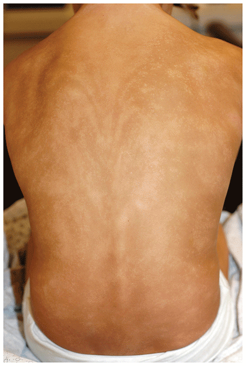
▶One pattern of mosaic hyperpigmentation affects 1 or several large regions or segments of the body and has been termed, in some cases, segmental pigmentation disorder (Figure 76.1). Another typical pattern is whorled or linear bands (thin or broad) that follow the Blaschko lines (Figures 76.2 and 76.3). In some cases, patients may have a mixture of hypopigmentation and hyperpigmentation, making it difficult to determine the typical background skin type (Figure 76.4).
▶Affected areas are typically sharply demarcated and often stop at the midline.
Figure 76.1. Pigmentary Mosaicism, Hyperpigmented Type. This Young Child Has a Large Hyperpigmented Patch Involving a Large Region of the Right Side of the Abdomen (Segmental Pigmentation Type).
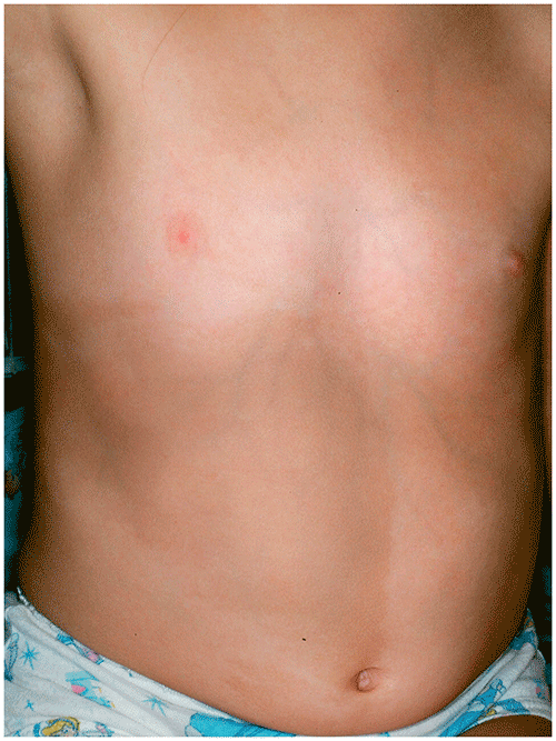
Figure 76.2. Pigmentary Mosaicism, Hyperpigmented Type. This Child Has Linear and Curvilinear Hyperpigmented Patches that Follow the Blaschko Lines, Limited to the Right Side of the Upper Back.
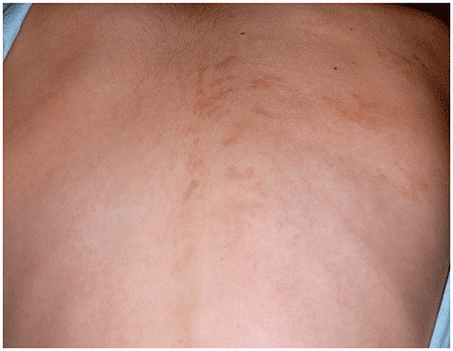
Figure 76.3. Pigmentary Mosaicism, Hyperpigmented Type. There is an Area of Hyperpigmentation Composed of Coalescent Shaggy-Bordered Macules and Patches.
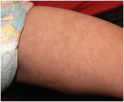
Figure 76.4. Pigmentary Mosaicism with Mixed Pattern of Hyperpigmentation and Hypopigmentation that Follows the Blaschko Lines on the Back.
Look-alikes
| Disorder | Differentiating Features |
|---|
| McCune-Albright syndrome | In addition to large café au lait patches with a “coast of Maine” (rough or shaggy-bordered) appearance (which may appear similar to segmental pigmentation type of pigmentary mosaicism), polyostotic fibrous dysplasia (often presenting as fractures) and endocrine hyperfunction (which may present as precocious puberty) are seen. Skin and bone changes are typically unilateral (although this is also the case with localized pigmentary mosaicism).
|
| Incontinentia pigmenti (third stage) | X-linked dominant inheritance. Vesicles are usually the initial presentation in newborn (first stage), distributed along the Blaschko lines. Warty lesions (second stage) give rise to eventual hyperpigmentation (third stage) in a similar distribution pattern. Associations may include dental, ophthalmologic, neurologic, and skeletal anomalies; irregular patches of scalp alopecia may also be present.
|
| Becker nevus (pilar and smooth muscle hamartoma) | An irregular hyperpigmented patch, plaque, or band occurring on the torso, often over the shoulder region. Often presents or enlarges around the time of puberty. Associated hypertrichosis and a pseudo-Darier sign (contraction of prominent arrector pili muscles) may be visible after stroking.
|
| Plexiform neurofibroma (in neurofibromatosis type 1) | In addition to hyperpigmentation, may have a “bag of worms” consistency at palpation. May have overlying hypertrichosis. Other signs of neurofibromatosis type 1 are present.
|
▶The diagnosis of hyperpigmented pigmentary mosaicism is usually made based on history and physical examination.
▶Consider a formal ophthalmologic examination to evaluate for ocular anomalies in children with the generalized type.
▶If other malformations or neurodevelopmental variations are absent, further workup is not indicated. If they are present, consultation with the appropriate specialists is warranted.
▶Rarely, karyotype analysis is performed (on blood or skin biopsy tissue) to search for chromosomal mosaicism.
▶In most cases, hyperpigmented pigmentary mosaicism is a benign, isolated skin finding not associated with other medical concerns.
▶In the rare patient who has generalized involvement, prognosis depends on the nature of any other organ variations.
▶Referral to dermatologic examination is warranted when diagnosis is unclear.
▶Referral to other specialists (eg, ophthalmology, neurology, genetics, orthopedics) is warranted when applicable.