▶Usually begins 24 to 48 hours after birth; rarely, lesions may be present at birth or appear as late as 10 days after birth.
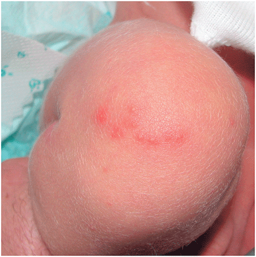
▶Appears as discrete, blotchy erythematous macules or patches, each with a central papule, vesicle, or pustule (Figures 103.1 and 103.2).
▶Occasionally, there may be clusters of papules, vesicles, or pustules that form an erythematous plaque.
▶Palms and soles are spared.
▶New lesions appear for several days; the process lasts a week or less.
Figure 103.1. Erythematous Macules, Each with a Central Papule, are Typical of Erythema Toxicum.
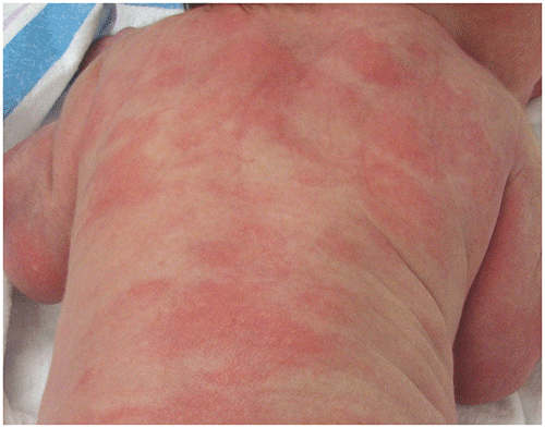
Figure 103.2. Erythematous Papules of Erythema Toxicum Located on the Knee.
Look-alikes (in descending order of frequency of occurrence)
| Disorder | Differentiating Features |
|---|
| Transient neonatal pustular melanosis | Most often seen in Black newborns; rare in other racial groups. Pustules (without erythema) or ruptured pustules that appear as small freckle-like hyperpigmented macules surrounded by a rim of scale. Pustular fluid contains neutrophils.
|
| Miliaria crystallina | |
| Neonatal acne (also termed neonatal cephalic pustulosis) | |
| Staphylococcal folliculitis | White to slightly yellow pustules with surrounding rim of erythema. Hair may be noted protruding centrally. Gram stain and bacterial culture will reveal Staphylococcus aureus.
|
| Bullous impetigo | Flaccid bullae or ruptured bullae forming round or oval crusted erosions; vesicles occasionally present. Gram stain and culture will reveal S aureus. Occasionally presents as tense inflammatory pustules (referred to as staphylococcal pustulosis); pustules larger than seen in erythema toxicum and blotchy erythema absent.
|
| Scabies | Occurs rarely during the first month after birth. Generalized eruption; may have vesicles but usually will be accompanied by erythematous papules or nodules and linear burrows. Palmoplantar involvement common. Mineral oil preparation of scrapings of papules will reveal mites, eggs, or fecal material.
|
| Neonatal herpes simplex virus infection | Typically, clustered vesicles on an erythematous base (although solitary vesicles occasionally occur). Lesions concentrated on the head, particularly at sites of trauma (eg, that caused by a scalp electrode). Neonates may have signs of sepsis (in disseminated disease) or seizures or coma (in central nervous system disease). Tzanck test, direct fluorescence examination, viral culture, or polymerase chain reaction (cerebrospinal fluid) will confirm diagnosis.
|
| Congenital candidiasis | Widespread rash composed of many tiny erythematous papules and pustules and scaling. Potassium hydroxide preparation of scale or a pustule roof will reveal pseudohyphae or spores. Palmoplantar involvement common. Nail changes (eg, yellow discoloration, ridging) may be present.
|
| Infantile acropustulosis | Usually begins in first months (not in first days) after birth. Vesicles or pustules limited to hands and feet, including palms, soles, wrists, and ankles. Episodes last 5 to 10 days and reappear every 2 to 4 weeks.
|
| Incontinentia pigmenti | |
| Eosinophilic pustular folliculitis | Papules and pustules, typically located on scalp. Exhibits chronic, intermittent course.
|
▶The diagnosis is made clinically. If uncertainty exists, use of a Wright stain of vesicular fluid will reveal a predominance of eosinophils.
▶Using a Tzanck smear, viral culture, direct fluorescence examination, polymerase chain reaction, Gram stain, or bacterial culture will assist in excluding infectious causes.
▶Skin biopsy rarely is required to exclude incontinentia pigmenti.
▶Obtain consultation if presentation is atypical (eg, suggesting an alternate diagnosis such as herpes simplex virus infection, incontinentia pigmenti).