▶A rare immunobullous disorder in children.
▶Associated with celiac disease (ie, gluten-sensitive enteropathy) in 75% to 95% of patients who are affected.
▶Typically seen in children between 2 and 7 years of age.
▶Children with celiac disease diagnosed have circulating immunoglobulin A antibodies to tissue transglutaminase and endomysium.
▶Characterized by intensely pruritic papulovesicular lesions (Figure 84.1) with a bilateral, symmetric distribution.
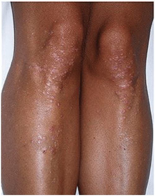
▶Most often located on the extensor aspects of knees and elbows, sacrum, buttocks, posterior aspect of the neck, scalp, and shoulders (Figures 84.2 and 84.3).
▶Mucous membrane involvement usually absent.
Figure 84.1. Dermatitis Herpetiformis. Vesicles and Erosions are Present.
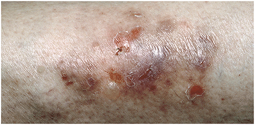
Figure 84.2. Intensely Pruritic Lesions of Dermatitis Herpetiformis on the Leg of a Patient with Celiac Disease.
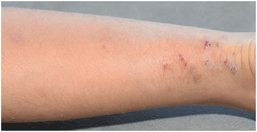
Figure 84.3. Grouped Papules, Vesicles, and Erosions on the Knees and Legs of a Patient with Skin of Color with Dermatitis Herpetiformis. Reproduced with Permission from Antiga E, Et Al. Dermatitis Herpetiformis: Novel Perspectives. Front Immunol. 2019;10.
Look-alikes
| Disorder | Differentiating Features |
|---|
| Linear immunoglobulin A dermatosis | Bullae tend to be larger. “Cluster (or string) of jewels” pattern (annular grouping of bullae) often noted. Not as symmetric in distribution, less often pruritic. Direct immunofluorescence and immunoblotting studies help confirm diagnosis.
|
| Bullous lupus erythematosus | Other features of systemic lupus erythematosus usually present. Concentrated in sun-exposed areas. Bullae tend to be larger, not typically pruritic. Antinuclear antibody (and other serological studies) will confirm diagnosis of systemic lupus erythematosus. Direct immunofluorescence and immunoblotting studies help confirm the diagnosis.
|
| Bullous pemphigoid | Urticarial plaques present in addition to tense blisters. Bullae tend to be larger. Pruritus common with early lesions. Direct immunofluorescence and immunoblotting studies help confirm the diagnosis.
|
| Herpes simplex virus infection | Most often clustered vesicles and erosions on an erythematous base. Often occur inside or around the mouth. Tend to be painful, less often pruritic. More often focal.
|
| Arthropod bites and papular urticaria | Papules may have a central punctum at close inspection. Usually concentrated on exposed areas of skin. Linear groupings of papules may be observed owing to exposure to crawling (and feeding) insects.
|
| Scabies | Mixture of papules, linear burrows, crusted papules or (especially in infants) nodules. Palm and sole lesions very common, as is involvement of the areolae and penis. Other family members often report lesions and/or pruritus. Microscopic examination of skin scrapings with mineral oil confirms the diagnosis (visualization of scabies mites, eggs, or fecal pellets). Pruritus worse in the evening.
|
| Pityriasis lichenoides et varioliformis acuta | Scaly, red papules with necrotic surface changes. Usually not pruritic. Associated fever may be present. Usually responds to treatment with oral erythromycin or tetracycline.
|
| Epidermolysis bullosa | Inherited (not acquired) mechanobullous disease. Usually begins in neonatal period or during early infancy. Vesicles and bullae induced by friction, pressure, or trauma. Blisters usually larger than those seen in dermatitis herpetiformis (with exception of some simplex forms of epidermolysis bullosa). Certain subtypes may reveal nail dystrophy, milia, extensive scarring, mitten deformities. Molecular genetic testing confirms the diagnosis; immunofluorescence antigen mapping and electron microscopy of skin biopsy specimens used less often in current era.
|
| Acquired epidermolysis bullosa | Acquired autoimmune blistering disease. Blisters induced by trauma. Scarring common. Direct fluorescence and immunoblotting studies will help confirm diagnosis. Blisters and erosions usually larger than those seen in dermatitis herpetiformis.
|
▶The diagnosis is suggested clinically and confirmed by means of skin biopsy with immunofluorescence study, which reveals immunoglobulin A at dermal papillary tips in a granular pattern.
▶Circulating serum antibodies to tissue transglutaminase or endomysium may be present (in patients sensitive to gluten).
▶Dapsone at 1 to 2 mg/kg/d is usually very effective (must first confirm appropriate glucose-6-phosphate dehydrogenase level and follow complete blood cell counts and liver function tests).
▶Sulfapyridine may be an effective alternative for therapy.
▶Gluten-free diet may be effective in certain patients, although challenging for children and parents.
▶Patients with gluten sensitivity should be referred to an experienced gastroenterologist for baseline and follow-up care.
▶The prognosis for children with dermatitis herpetiformis is unpredictable.
▶Many recommend indefinite continuation of the gluten-free diet in individuals sensitive to gluten, as even small amounts of gluten exposure can lead to disease relapse.