▶Papulosquamous (ie, elevated lesions with scale) condition with a tendency to persist or recur for years.
▶Characterized by inflammation and hyperproliferation of the epidermis.
▶Likely results from both a genetic predisposition (family history often is positive) and an environmental trigger (eg, stress, infection, trauma).
▶Recent literature links psoriasis to other systemic comorbidities, in particular a higher prevalence of metabolic syndrome (ie, obesity, dyslipidemia, hypertension, and elevated blood glucose levels) and cardiovascular disease in children and adults. Psoriasis may occur in some children with juvenile idiopathic arthritis. Typically, this occurs within 2 years of diagnosis of juvenile idiopathic arthritis.
▶Appearance of lesions
■Lesions are well-defined papules, patches, and plaques that are pink to deep red and have an adherent white to silvery “micaceous” scale (Figure 52.1).
– Removal of scale produces pinpoint bleeding points (Auspitz sign) due to dilated dermal capillaries (Figure 52.2).
– Scale may be absent or less prominent in moist, intertriginous areas (eg, diaper area, axillae) (Figure 52.3).
■Variants
– Infantile psoriasis: may appear as generalized erythroderma or as sharply demarcated erythema (with minimal scale) in diaper region (Figure 52.4), axillae, and umbilicus.
– Guttate psoriasis: Often precipitated by pharyngeal or perianal Streptococcus pyogenes infection; begins as generalized erythematous macules and papules (that may mimic a viral exanthem); later, characteristic scale appears (Figure 52.5).
– Pustular psoriasis: small pustules studded over the surface of deep red plaques (Figure 52.6).
– Inverse psoriasis: lesions located predominantly in the axillae (Figure 52.7) and groin.
▶Distribution
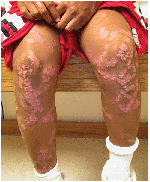
■Scalp (scaling and erythema) (Figure 52.8), posterior auricular regions, elbows (Figure 52.9), knees (Figure 52.10), umbilicus, and gluteal cleft; however, any body region may be affected.
■Lesions appear in areas of trauma (ie, Koebner phenomenon), explaining involvement of the extensor surfaces of the extremities.
■Nail involvement is common, consisting of pitting or thickening and yellowing.
Figure 52.1. Typical Lesions of Psoriasis are Erythematous Papules and Plaques that Have a Thick Adherent Scale.
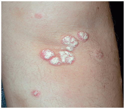
Figure 52.2. Plaque of Psoriasis with Auspitz Sign (Pinpoint Bleeding in Areas of Removed Scale).
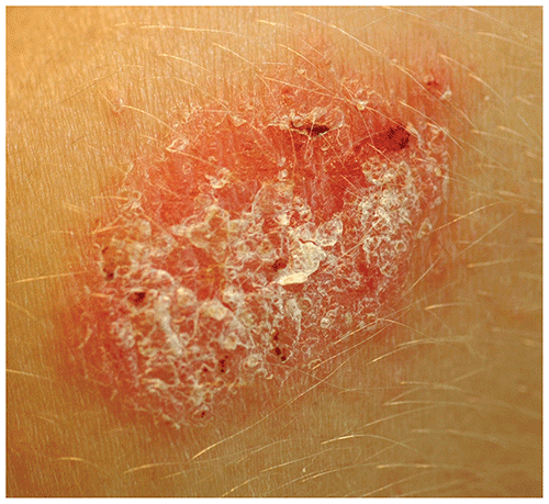
Figure 52.3. In Occluded Areas, Such as the Axilla, the Lesions of Psoriasis May Lack Scale.
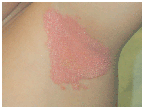
Figure 52.4. In Infants, Psoriasis May Involve the Diaper Area, Appearing as Sharply Defined Erythematous Patches with Little Scale. Also Note Involvement of the Umbilicus, a Common Finding in Psoriasis.
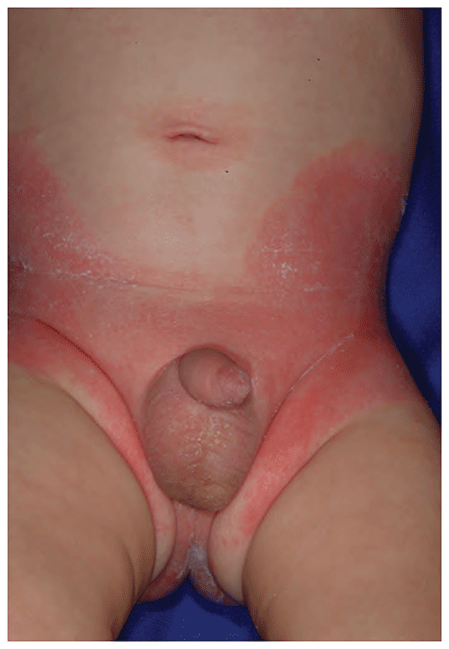
Figure 52.5. Guttate Psoriasis is Characterized by an Eruption Composed of Widespread Macules or Papules that May Mimic a Viral Exanthem. Over Time, the Lesions Develop Thick Scale.
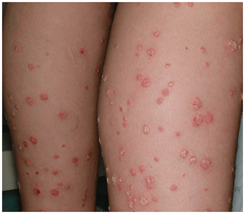
Figure 52.6. Pustular Psoriasis. This Patient Presented with Numerous Pustules Overlying Red Plaques.
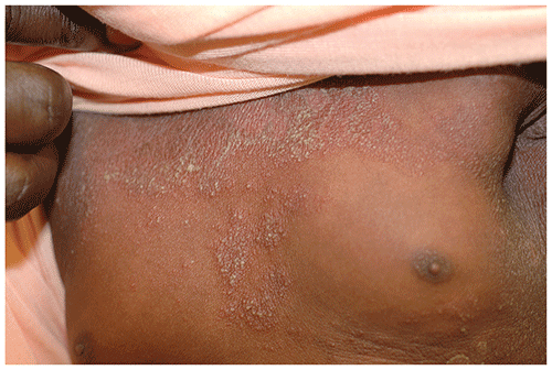
Figure 52.7. Left Axillary Vault with a Plaque of Inverse Psoriasis (Note Reduced Scaling Due to Intertriginous Location).
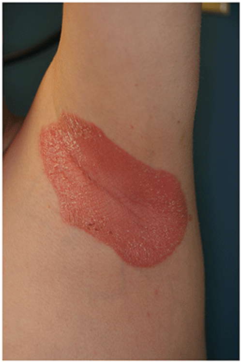
Figure 52.8. On the Scalp, Psoriasis Causes Erythema and Thick Scale.
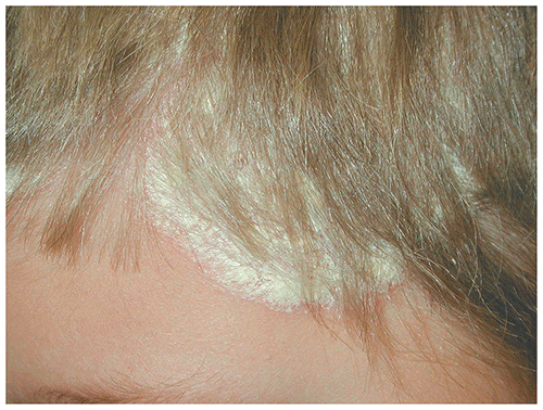
Figure 52.9. Psoriatic Papules Coalescing into Plaques on the Elbows of an Affected Teen.
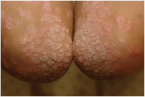
Figure 52.10. Patient with Psoriasis of the Knees and Shins.
Look-alikes
| Disorder | Differentiating Features |
|---|
| Lichen planus | Purple or pink polygonal papules and small plaques. Fine scale (not thick adherent scale) is present. Oral involvement common, with thin, white, elevated linear lesions forming a lacy, reticulated appearance.
|
| Dermatomyositis | Patients may exhibit muscle weakness. Characteristic cutaneous findings include heliotrope rash and Gottron papules (ie, erythematous papules located over the dorsal surfaces of interphalangeal joints of the fingers). Lesional scaling is typically minimal.
|
| Pityriasis rosea | May be confused with guttate psoriasis. Many patients first develop a herald patch followed by small, thin oval plaques with long axes parallel to lines of skin tension. Lesions covered by thin, fine ”trailing scale” (lags behind advancing red border, and free edge of scale points inward toward center of plaque).
|
| Seborrheic dermatitis | May be difficult to distinguish from psoriasis when only the scalp and face are involved. Typical lesions have greasy, yellow/tan scales. Auspitz sign absent. In infants, seborrheic dermatitis and psoriasis may be indistinguishable.
|
▶The diagnosis is made clinically based on the appearance and distribution of lesions.
▶Observation of Auspitz sign strongly suggests a diagnosis of psoriasis.
▶Skin biopsy can be helpful if the clinical presentation is not diagnostic.
▶Therapy is directed at reducing inflammation and normalizing epidermal proliferation.
▶Topical therapy (first line): Treatment often involves more than 1 agent.
■Mid-potency (or, occasionally, high-potency) topical glucocorticoids (often used in conjunction with calcipotriene).
■Calcipotriene (vitamin D derivative): Normalizes epidermal proliferation; may be used as monotherapy or in conjunction with topical corticosteroids; may cause hypercalcemia in infants or small children if applied over large surface areas.
■Others: anthralin or liquor carbonis detergens (also known as LCD, a tar derivative), topical phosphodiesterase-4 inhibitors, and topical retinoids occasionally used.
■Moisturization is recommended to help maintain skin integrity.
▶Phototherapy or photochemotherapy: UV-B or UV-A (alone or combined with psoralen, when known as PUVA) therapy may be used for patients with severe disease in whom topical therapy fails.
▶Systemic therapy: Methotrexate, cyclosporine A, or acitretin may be used for patients with severe disease in whom topical therapy fails. Biologic agents (that target tumor necrosis factor-α, interleukin [IL]-12, IL-17, and/or IL-23) are increasingly used, with pediatric approval (for psoriasis) down to 6 years of age for ustekinumab, secukinumab, and ixekizumab and down to 4 years of age for etanercept.
▶When guttate psoriasis is suspected, test for pharyngeal (or perianal if the physical examination is suggestive) S pyogenes infection and treat if infection is confirmed.
▶Psoriasis is a chronic disease, and recurrences may be anticipated.
▶For patients with significant disease, consult with or refer to a pediatric dermatologist to optimize therapy.
▶Refer patients in whom the diagnosis is uncertain, those in whom appropriate therapy does not work, or those in whom pustular disease develops.
▶If there is clinical concern for possible psoriatic arthritis, refer to a pediatric rheumatologist.