▶Also referred to as localized scleroderma, morphea is an uncommon autoimmune inflammatory sclerosing disorder of the skin and subcutaneous tissue.
▶While the term scleroderma and the characteristic hardening (sclerosis) of the skin can be confused with systemic sclerosis, morphea does not progress to systemic disease. Rarely, though, morphea can coexist with systemic sclerosis. While it is variable, some clinicians prefer the term morphea over localized scleroderma.
▶The occurrence is estimated to be 0.4 to 1 per 100,000 individuals, and the condition is 2 to 3 times more common in female than male patients; recent studies suggest morphea is more prevalent in white female patients.
▶Mean age of onset in children is 5 to 8 years; however, morphea has been described in infants, and there are even case reports of congenital morphea.
▶While most adults have disease limited to the skin and subcutaneous tissue, children have a higher risk for extra-cutaneous findings.
▶Divided into several subtypes; clinical features vary depending on the subtype of morphea.
▶Disease severity ranges from a solitary area of induration (hardening of skin) to severe, disfiguring and disabling disease affecting skin, subcutaneous tissue, underlying muscle and bone, and brain, ocular, and oral tissues. Involvement of the lung, heart, and gastrointestinal tract can also occur, but unlike with systemic sclerosis, this involvement is usually mild and nonprogressive.
▶Occasionally observed in the setting of other connective tissue disorders, including juvenile idiopathic arthritis, systemic lupus erythematosus, systemic sclerosis, Sjögren syndrome, juvenile dermatomyositis, polymyositis, and eosinophilic fasciitis.
Skin Findings
▶Begins insidiously as a gradual hardening of the skin and/or a change in skin color (eg, erythematous, violaceous).
▶May begin with an inflammatory stage that presents with erythema or a violaceous discoloration of the skin (Figure 119.1); at times, this can resemble a port-wine stain (capillary malformation).
▶With time, the redness fades and the affected area of skin becomes indurated and often ivory-colored and shiny in appearance (Figure 119.2).
▶Over time, lesions become hyperpigmented and/or atrophic (see Figures 119.1 and 119.3). These changes usually evolve gradually over several months and are seen in older “burnt-out” lesions. However, these signs may coexist with active disease, so their presence does not completely exclude ongoing disease activity.
▶Lesions can spontaneously soften over time.
▶Overall clinical appearance varies depending on the subtype. There is no universally agreed-on classification of morphea, but a pediatric proposed classification system divides morphea into 5 subtypes:
■Linear morphea (also called linear scleroderma)
■Circumscribed (plaque) morphea
■Generalized morphea
■Pansclerotic morphea
■Mixed morphea
▶Linear morphea is the most common type of morphea in children and most often affects the face and/or extremities.
■Hardening of the skin or subcutaneous tissue (as well as the associated hypopigmentation or hyperpigmentation) spreads in a linear distribution, most commonly over an extremity (Figure 119.4) or on the face (Figure 119.5). The distribution follows an embryonic pattern known as Blaschko lines.
■Early findings of erythema and violaceous discoloration are more subtle in linear versus circumscribed (plaque) morphea; the early erythema can occasionally be confused with a port-wine stain.
■When involving the forehead and scalp, referred to as en coup de sabre (cut of a saber) (Figures 119.6 and 119.7). Tends to have a unilateral distribution in most children; the skin gradually becomes more atrophic and develops a depressed, groove-like appearance. Can also affect underlying bone, causing disturbances to typical growth.
■More extensive hemifacial involvement may occur; it is termed progressive facial hemiatrophy (Parry-Romberg syndrome).
■Scalp involvement may be associated with alopecia (see Figure 119.6); can also spread inferiorly to involve the periorbital region (Figure 119.8), nose, and mouth; may become quite impacted as a result of marked atrophy.
■Morphea may be associated with significant atrophy of the skin and subcutaneous tissue; when involving a limb, this can result in circumferential and linear undergrowth of the affected extremity (Figures 119.9 and 119.10). Untreated, these problems can result in the need for orthopedic corrective surgery, which has included amputations in the past.
■When extending over a joint, morphea may result in contractures and impaired mobility or range of motion, which can be permanent.
▶Circumscribed (plaque) morphea is the second most common subtype of morphea in children.
■May present with one or more plaques of affected skin, most often located on the trunk. Affected skin usually begins with an oval or round circumscribed area of induration.
■Often begins with erythematous or violaceous discoloration (see Figure 119.1), which gradually evolves to the characteristic ivory color with increasing induration; as the process progresses, the erythema or violet hue fades.
■Circumscribed morphea can be further categorized as superficial or deep, depending on the depth of skin and soft-tissue involvement.
▶Generalized morphea is defined by some experts as multiple plaques of morphea covering at least 30% of the body.
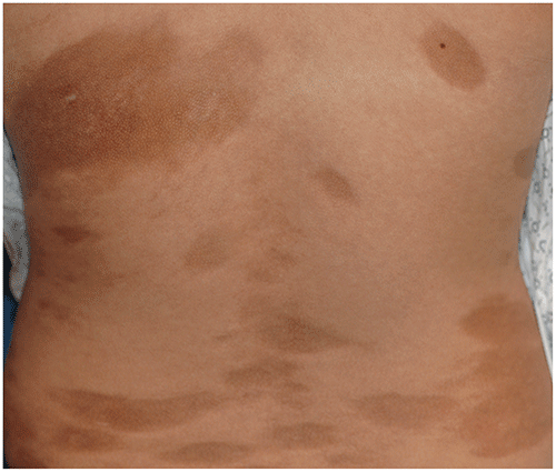
■Classification defines generalized morphea as 4 or more individual plaques measuring more than 3 cm (which may become confluent), involving at least 2 of 7 anatomic sites (ie, head and neck, right or left arm, right or left leg, anterior trunk, posterior trunk) (Figure 119.11).
▶Pansclerotic morphea is a rare subtype involving the skin, subcutaneous tissue, muscle, and bone, often on an extremity.
■Usually results in circumferential involvement with significant cosmetic and functional compromise; skin may show pitting edema or diffuse painful areas with a puckered or peau d’orange texture.
– This subtype can be life-threatening when rapidly progressive, as it can lead to widespread involvement with skin breakdown that not only puts the patient at risk for sepsis but also increases the risk for squamous cell cancer.
▶Mixed morphea.
■Some patients do not clearly have any of the subtypes of morphea because they have features of more than one subtype, most commonly a combination of linear and circumscribed (plaque) morphea.
Figure 119.1. Circumscribed (Plaque) Morphea. There is a New Lesion Shown in the Center of the Photograph. It is an Erythematous Patch with a More Intensely Erythematous to Violaceous Border. Resolving Lesions are Seen as Hyperpigmented Patches.
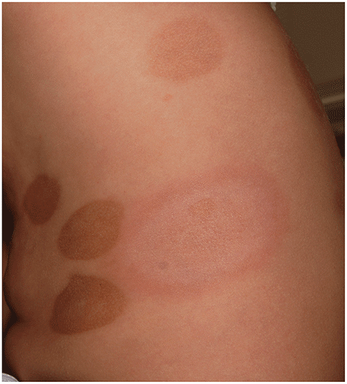
Figure 119.2. Morphea of the Thigh. An Ivory-Colored Indurated Plaque with Surrounding Peripheral Erythema.
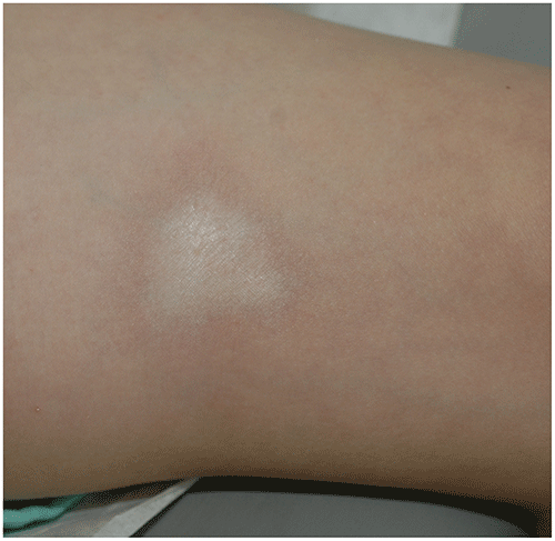
Figure 119.3. Localized Morphea on the Back of a Teenager. This Older Lesion is Characterized by an Atrophic Plaque.
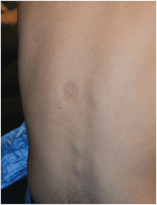
Figure 119.4. Linear Morphea Involving the Arm. The Lesions are Ivory-Colored and Indurated. Hyperpigmentation is Developing.
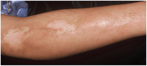
Figure 119.5. Linear Morphea Involving the Face. Note the Atrophy Affecting the Chin to the Left of the Midline.
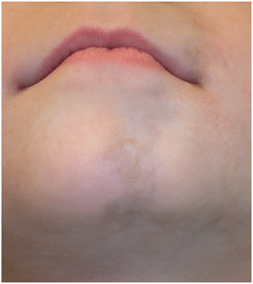
Figure 119.6. En Coup De Sabre (Cut of a Saber) Form of Linear Morphea. There is a Linear Area of Atrophy and Alopecia Involving the Frontal Scalp.
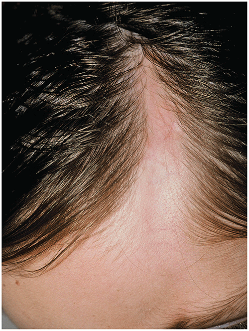
Figure 119.7. In This Child with the En Coup De Sabre (Cut of a Saber) Form of Linear Morphea, Resolving Lesions Have Become Hyperpigmented.
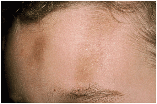
Figure 119.8. Patient with Periorbital Morphea in Early Stages.
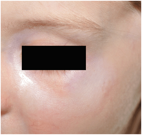
Figure 119.9. This 5-Year-Old Had Extensive Morphea of the Lower Extremity, Resulting in Circumferential and Linear Size Discrepancy with the Unaffected Side.
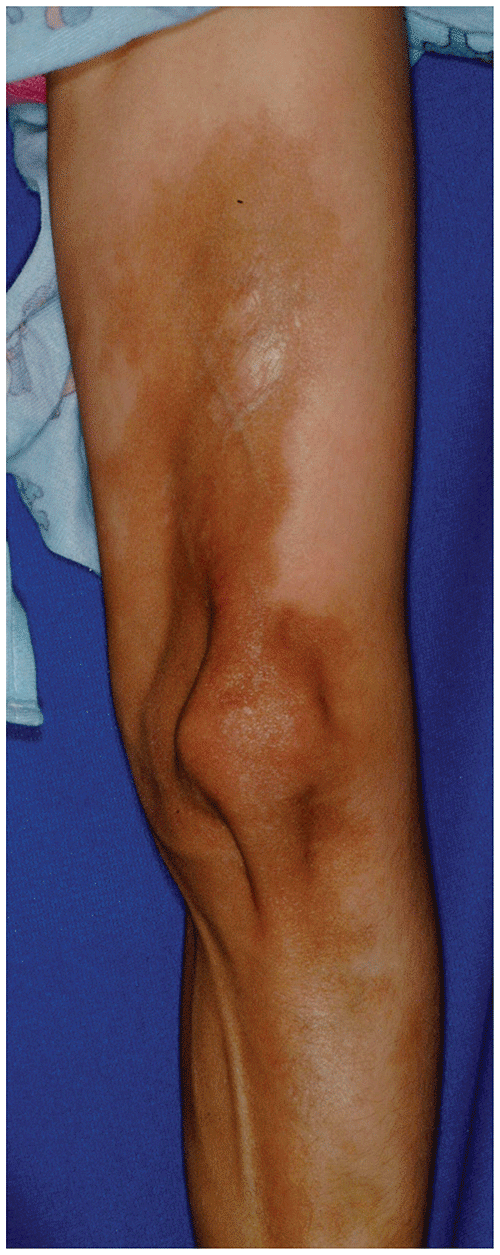
Figure 119.10. Patient with Morphea of the Left Leg with Leg Length Discrepancy and Left Foot Hypoplasia.
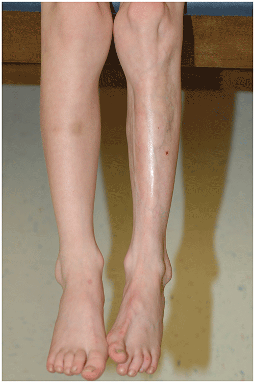
Figure 119.11. Generalized Morphea. Multiple Lesions Involving the Trunk Have Become Hyperpigmented, and Many are Atrophic.
Other Clinical Signs
It has become increasingly apparent that many patients with morphea experience associated extra-cutaneous involvement. A retrospective, multicenter international study of 750 patients with juvenile localized scleroderma (morphea) revealed that nearly one-quarter had extra-cutaneous features, while a more recent prospective, multicenter North American study of 86 patients identified a much higher extra-cutaneous frequency of 57%. Another study concluded that the risk for extra-cutaneous symptoms was greater among patients with disease onset before 10 years of age.
▶Musculoskeletal findings are the most common type of extra-cutaneous problem, including contractures, arthralgias, arthritis, myalgia, myositis, weakness, and atrophy. Bony undergrowth has also been identified in about a quarter of the patients. Musculoskeletal manifestations are more common in those with linear scleroderma. Arthritis may affect joints with overlying skin lesions but also occurs remotely from the skin involvement in approximately 25% of patients.
▶Neurologic problems have been reported, primarily with linear morphea of the upper face. Central nervous system manifestations include headaches, seizures, peripheral neuropathy, and behavioral or learning differences. Seizures can be complex and very difficult to control. Headaches appear to be the most common neurologic problem, and in some patients, they present as migraines, including status migrainosus. Changes seen at magnetic resonance imaging, including white matter lesions, intracerebral atrophy, and calcifications, have been reported in some patients. Rarely, vasculitis or vasculopathy has been found on brain biopsy specimens.
▶Ocular manifestations may include episcleritis, uveitis, keratitis, xerophthalmia, glaucoma, and papilledema. Ophthalmologic evaluation is recommended for all patients with morphea at diagnosis, with routine follow-up screening recommended for those with craniofacial involvement.
▶Vascular involvement (most commonly Raynaud phenomenon), gastroesophageal reflux or dysphagia, and involvement of the cardiac, pulmonary, and renal systems are infrequently associated with morphea. These problems are typically mild and nonprogressive, unlike the pattern found in systemic sclerosis.
▶Extra-cutaneous involvement has been associated with more symptoms in the child (eg, trouble playing), poorer response to treatment (need for more treatments/longer treatment courses), and greater disease effects.
▶Psychologic implications and functional impairment may result from this chronic disease. Outcome is improved by earlier treatment initiation, with use of systemic immunosuppressants recommended for patients with active disease who are at risk of experiencing poor outcome.
Look-alikes
| Disorder | Differentiating Features |
|---|
| Capillary malformation (port-wine stain) | May resemble early inflammatory stage of morphea. Usually present at birth, unlike most morphea. Usually stable during first several years after birth. Flat and smooth, not firm or indurated, on palpation. Some capillary malformations are associated with overgrowth (but not typically atrophy) of underlying tissues.
|
| Lichen striatus | May resemble early lesions of linear morphea. Lichenoid skin-colored to pink papules that coalesce in a linear/band-like pattern; follows the lines of Blaschko. No firmness or induration of affected skin. Self-limited condition that often resolves spontaneously over 1–2 years. May leave persistent hypopigmentation or hyperpigmentation, but no atrophy of affected skin.
|
| Lichen sclerosus et atrophicus (LSA) | May also show sclerotic white plaques with atrophy. Most often involves the genitalia. More likely to be associated with severe pruritus than morphea. Extragenital LSA more likely to be associated with skin dryness than morphea. Sclerotic white scar-like lesions usually smaller than those of morphea; may have guttate (“teardrop”) pattern. More likely to see telangiectasias or follicular plugging in LSA than with morphea. Hemorrhagic blisters occasionally present.
|
| Pasini-Pierini atrophoderma | Considered by some clinicians to be a superficial variant of morphea. Hyperpigmented (brown to gray to blue) patches seen most commonly on the back. Lesions lack induration; preceding inflammatory phase always absent. Face, hands, and feet usually spared. Lesion borders are sharply defined; described as having “cliff-drop” borders ranging from 1–8 mm in depth. Duration of many years to decades with benign course.
|
| Linear atrophoderma of Moulin | Atrophic plaques that are linear and may follow the lines of Blaschko. Inflammation, induration, and pigmentary changes all absent.
|
| Acrodermatitis chronica atrophicans | Cutaneous manifestation of chronic Lyme disease. Violaceous plaques of the distal extensor extremities that may become indurated, hyperpigmented, and atrophic. Primarily seen in Europe, linked to Borrelia infection (connection between Borrelia infection and morphea remains controversial). May have associated arthritis and neuropathy. Primarily in adult women, occurring several months to years after initial infection. Lyme antibody titer results may be positive.
|
| Systemic sclerosis (scleroderma) | Generalized disorder that may affect many organs in addition to the skin: lungs, kidneys, heart, gastrointestinal tract, and/or joints. Much less common in children than adults. Very rare when younger than 5 years. Nail fold capillary changes found in nearly all patients similar to those seen in juvenile dermatomyositis. Early signs include Raynaud phenomenon, poor weight gain or weight loss, arthralgia, myalgia, and fatigue. Raynaud phenomenon can precede systemic sclerosis development by months to years. Often associated with characteristic facial features: pinched nose, pursed lips, and small oral aperture. Puffiness of the fingers (induration) is followed by sclerodactyly (shiny tapered fingertips with limited range of motion), with hand involvement found in most patients. Other common features include arthralgias, decreased joint mobility, weight loss, fatigue, gastrointestinal symptoms, and shortness of breath with exertion. Telangiectasias, calcification, and ulceration of the skin may occur (rarely seen in morphea). Skin involvement is bilateral, symmetric, and diffuse, different from the linear or circumscribed lesions of morphea.
|
| Chronic graft-versus-host disease, sclerodermatous type | History of preceding at-risk procedure (ie, bone marrow or stem cell transplant) and immunosuppression in host. Widespread sclerodermatous plaques may be seen, similar to those of systemic sclerosis or generalized morphea. May be associated with cutaneous ulceration, nail dystrophy, scarring alopecia, and joint contractures. Erosions of the oral mucous membranes often present. Systemic manifestations may include gastrointestinal, hepatic, pulmonary, cardiac, or hematologic aberrations. Skin biopsy usually shows interface dermatitis in addition to dermal sclerosis.
|
| Eosinophilic fasciitis | Generalized infiltration/induration of skin of the trunk and/or extremities; classically spares hands, feet, and face (although hands and feet occasionally involved). Abrupt onset of painful skin swelling. Cobblestoned or puckered appearance of the skin may be present. Usually responds well to systemic steroids. Usually not well circumscribed or linear in distribution. Associated with striking peripheral eosinophilia (but may rapidly correct on administration of systemic steroids), elevated erythrocyte sedimentation rate, and hypergammaglobulinemia.
|
| Nephrogenic systemic fibrosis (nephrogenic fibrosing dermopathy) | Usually seen in patients with renal insufficiency and exposure to gadolinium-based contrast media. Often associated with a hypercoagulable state. Poorly defined, indurated plaques usually distributed symmetrically on the extremities. Frequently associated with joint contractures, pain, and decreased mobility. May develop fibrosis of the heart, lungs, and skeletal muscle.
|
| Progeria | Very rare premature aging syndrome. Diagnosis should be considered in young infants who present with widespread sclerodermatous plaques. Other characteristic features include thin and beaked nose, midfacial duskiness and hypoplasia, micrognathia, and slow growth. Prominent skin vasculature, especially over the scalp, develops over time. Small face with birdlike appearance. High-pitched voice. Prominent eyes with incomplete closure of eyelids. Caused by variation in LMNA gene.
|
▶In most children, the diagnosis of morphea is based on clinical features. Skin biopsy can be helpful to confirm a clinical diagnosis when uncertain.
▶Blood analyses may be used to screen for associated systemic autoimmune or rheumatologic disease but are not helpful in diagnosing morphea. In some patients, they may help with monitoring disease activity.
▶Patients with rapidly progressive skin disease, more extensive skin or deeper tissue disease, or extra-cutaneous involvement or risk for extra-cutaneous involvement (eg, craniofacial involvement, all subtypes except for circumscribed superficial morphea) would benefit from comprehensive physical and laboratory evaluation by a pediatric rheumatologist.
▶Patients with morphea are usually treated by a pediatric dermatologist and/or pediatric rheumatologist; collaborative care between both specialists is optimal and recommended if possible.
▶Clinical follow-up is challenging, and there is no consistently used tool to measure improvement or deterioration in disease; photographic comparisons, ultrasonography, thermography, and other imaging have all been used with variable consistency and utility; the Localized Scleroderma Cutaneous Assessment Tool (LoSCAT) and modified LoSCAT have been found useful in evaluating patient disease progression and in conducting clinical trials. However, LoSCAT assesses only skin involvement.
▶Damage and extra-cutaneous involvement occur early, with children at risk of developing a serious problem because of the early onset of the disease and long duration. The best strategy to minimize severe damage is through early identification and treatment initiation.
▶Treatment modality depends primarily on the severity of the patient’s morphea and may vary from close clinical observation to multidisciplinary management with the use of topical and systemic agents.
■Patients with mild, localized superficial morphea without extra-cutaneous involvement are most frequently treated with topical corticosteroids, calcipotriene (vitamin D analogue), or topical calcineurin inhibitors (tacrolimus ointment or pimecrolimus cream).
■Patients with deeper, more extensive disease with or at risk for extracutaneous involvement are treated with systemic immunosuppressive therapies, most commonly methotrexate with or without oral or intravenous corticosteroid pulse therapy. Mycophenolate mofetil is the next most commonly used systemic immunosuppressant. Several other treatments have been reported, including abatacept, tocilizumab, rituximab, Janus kinase inhibitors, and hydroxychloroquine, with combination therapy often used to control treatment-resistant disease.
■Phototherapy and photochemotherapy (particularly psoralen plus light from UV-A or UV-A1) has been used with success in the treatment of morphea, especially in adults. There is less experience with light therapy in the treatment of childhood morphea.
▶Careful history and physical examination should be performed in all patients with morphea to screen for associated clinical symptoms or physical findings. While morphea is confined to the skin and subcutaneous tissues in many patients, children are at increased risk for deep tissue and severe extracutaneous manifestations compared to those with adult-onset disease.
▶Extra-cutaneous involvement may be musculoskeletal, neurologic (primarily in patients with en coup de sabre distribution), ocular, oral, and, less commonly, vascular, gastrointestinal, pulmonary, or of another internal organ system. Early identification of deep tissue and extra-cutaneous tissue involvement is critical for reducing the risk for poor outcome, so early evaluation by a pediatric rheumatologist is recommended. Patients should be treated as necessary by a pediatric rheumatologist, pediatric dermatologist, pediatric ophthalmologist, or another appropriate subspecialist depending on the nature and extent of involvement.
▶Physical and occupational therapy referrals should be initiated as indicated.
▶Most adults with morphea have active disease for 3 to 5 years, but children have been found to have longer disease durations (13–13.5 years). Children also experience a higher relapse frequency than adults. Unlike with systemic sclerosis, most patients will go into spontaneous remission. The goal of treatment is to control activity to minimize damage, thereby reducing the risk for psychosocial and functional impairment and effects on quality of life.
▶A minority of affected children may continue to have morphea during adulthood, and another subset may experience relapse of active disease even after several years of remission.
▶Patients with limited plaque morphea usually do well with spontaneous remission after several years of disease activity. However, lesions of morphea can leave permanent pigmentary changes and atrophy of the affected skin, even after the active disease subsides.
▶Severe linear morphea can result in limited range of motion and atrophy of affected extremities, resulting in discrepancies in length and circumference between affected and unaffected limbs.
▶Children suspected of having cutaneous manifestations of morphea should be referred to a dermatologist or pediatric dermatologist for confirmation of the diagnosis and potential treatment.
▶Patients with facial involvement, widespread skin lesions, aggressive disease progression, pansclerotic morphea, linear scleroderma, and mixed morphea should be referred to a pediatric rheumatologist to be evaluated for extra-cutaneous involvement and the need for systemic immunomodulators.
▶Patients with severe morphea are often co-treated by pediatric dermatology and rheumatology specialists.