▶Epithelial growths are induced by different subtypes of human papillomavirus (HPV).
▶Clinical wart subtypes correlate with different HPV subtypes.
▶Very common in children.
▶Most spontaneously resolve over years.
▶Often recalcitrant to multiple therapies.
▶Transmission may occur person to person, from fomites, or from autoinoculation.
▶Usually asymptomatic, but large or multiple plantar lesions may be associated with pain, limitation in activities.
▶Can be disfiguring.
▶In patients who have immunodeficiency (including HIV infection), lesions may be numerous and widespread.
▶Common warts: discrete, skin-colored to hyperpigmented papules with characteristic verrucous surface (Figure 13.1). Sometimes, the surface is more mammillated (bumpy) (Figure 13.2). Lesions may exhibit tiny dark specks that are thrombosed capillaries. Some warts may have a filiform (stalklike) appearance (Figure 13.3).
▶Plantar warts: rough or smooth papules and plaques localized to the plantar aspect of the feet, most often over weight-bearing surfaces. Lesions exhibit tiny dark specks that are thrombosed capillaries. Occasionally, several warts may coalesce and form larger mosaic warts (Figure 13.4).
▶Flat warts: smooth, pink or skin-colored, flat-topped papules, 1 to 3 mm, typically seen on the face or legs, but may occur in other locations (Figure 13.5).
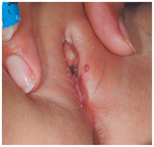
▶Anogenital warts (ie, condylomata acuminata): discrete papules or confluent plaques; pink to red or skin colored; localized to genitalia or adjacent skin of inguinal, thigh, suprapubic, or perianal areas (Figure 13.6).
▶Periungual warts: often occur in association with common warts; present as papules, confluent plaques, or nodules adjacent to nails, occasionally with destructive involvement of the proximal or lateral nail fold areas.
Figure 13.1. Common Warts Appear as Rough (Ie, Verrucous) Papules.
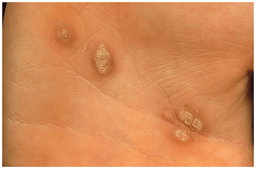
Figure 13.2. This Child with Skin of Color Had Postauricular Warts that Were Hyperpigmented, Mammillated (Rounded) Papules.
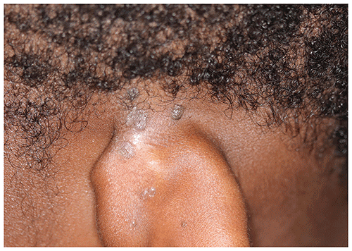
Figure 13.3. Verrucous Papules with Filiform (Stalklike) Appearance on the Medial Nares. Note the Smaller Smooth Papules on the Alar Rims, Which Also Were Warts.
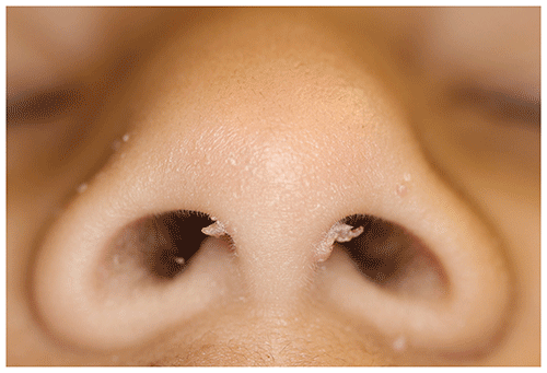
Figure 13.4. Multiple Verrucous Papules with Pinpoint Thrombosed Vessels Coalescing to Form Mosaic Plantar Warts. There is Mild Maceration Surrounding Some of the Lesions and Partial Clearance Due to Treatment with Salicylic Acid.
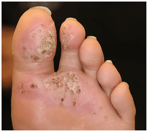
Figure 13.5. Flat Warts are Small, Flat-Topped Papules.
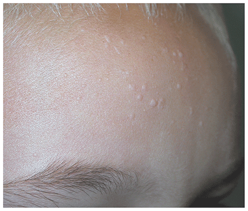
Figure 13.6. Condylomata Acuminata Appear as Skin-Colored Papules and Plaques.
Look-alikes
| Disorder | Differentiating Features |
|---|
| Plantar Warts |
|---|
| Callus | Located over points of friction or pressure. Lacks black specks (ie, thrombosed capillaries). Dermatoglyphics often preserved.
|
| Condylomata Acuminata |
|---|
| Condylomata lata | |
| Molluscum contagiosum | |
| Flat Warts |
|---|
| Lichen planus | |
| Lichen nitidus | |
| Molluscum contagiosum | |
| Benign cephalic histiocytosis | |
| Common Warts |
|---|
| Epidermal nevi | |
| Granuloma annulare | |
| Knuckle pads | Fibrous to hyperkeratotic plaques or papules overlying interphalangeal joints. Rough (ie, verrucous) surface tends to be absent.
|
▶Warts are often self-limited, usually asymptomatic, and do not necessarily require treatment. None of the current treatments are uniformly effective, and patients and parents should understand the potential limitations of therapy.
▶The risk-to-benefit ratio of therapy must be considered, and care should be exercised to avoid overly painful or traumatic treatments in young children.
▶First-line therapy is usually a topical salicylic acid plaster or liquid, with or without duct tape occlusion (Box 13.1).
▶A compounded cream of 5-fluorouracil and salicylic acid applied under tape occlusion nightly may be effective.
▶Cryotherapy with spray or cotton swab application of liquid nitrogen or other cryogen is effective if used repeatedly (with treatments separated by 2–4 weeks) but should be reserved for motivated, older children who can tolerate painful procedures. In the interval between cryotherapy treatments, any remaining wart should be treated with topical salicylic acid as described previously.
▶Topical imiquimod is approved for treatment of condylomata acuminata and is applied to lesions 3 times weekly on nonconsecutive days (and occasionally nightly) until resolved for up to 16 weeks.
▶Off-label use of imiquimod may be beneficial for common warts when applied daily to warts; however, its efficacy is often limited by the hyperkeratosis found in common warts, and irritant dermatitis may be seen with its use.
▶Cimetidine (30–40 mg/kg/d orally divided twice a day or 3 times a day) for 6 to 8 weeks or more may be effective in some children; not approved by the US Food and Drug Administration for this indication.
▶Other treatment options for common warts include intralesional injection of skin test antigens (eg, Candida, Trichophyton), intralesional chemotherapy injections (eg, bleomycin), and topical immunotherapy with squaric acid; these therapies are not approved by the US Food and Drug Administration, and published data are limited.
▶Other potential treatment options for condylomata acuminata include prescription therapies such as podofilox solution or gel, sinecatechins ointment, or in-office application of podophyllin or trichloroacetic acid.
▶Treatments such as pulsed dye laser and surgical excision are occasionally considered but do not necessarily offer greater efficacy. Surgery entails a high risk of permanent scarring and potential for recurrence.
Box 13.1. Optimizing Use of Over-the-Counter Salicylic Acid Therapy for Warts
| Apply 17% salicylic acid liquid to wart(s).
| | Air-dry for 2 to 3 minutes (develops into a white film). | | Occlude surface of wart with duct tape or similar adhesive tape. | | Remove tape in the morning. | | If further debridement is necessary, gently file tissue down with a disposable emery board to avoid autoinoculation. | | Repeat nightly until wart is resolved. | Notes
This treatment is most effective for plantar warts. Do not apply to facial, fold, or genital area warts. If area becomes macerated or inflamed, withhold treatment for 1 to 3 nights, then resume. May take up to 8 weeks to see improvement in the wart. Prolonged treatment is almost always necessary, particularly for plantar warts.
|
|
▶Patients with symptomatic warts that have not responded to standard therapies should be referred for discussion of other treatment options.
▶Patients who are immunosuppressed and have multiple lesions merit more aggressive therapy given the potential association between warts and an increased risk of cutaneous malignancy, as well as an increased likelihood of a more extensive and protracted course.
▶Anogenital warts in children may be a marker for sexual abuse, although autoinoculation, vertical transmission (a consideration primarily in children <3 years), and benign (nonsexual) modes of transmission are also possible. If the history or physical examination raises concern, referral and thorough investigation are vital.