▶Epidermolysis bullosa (EB) is a group of rare genetic skin disorders characterized by fragile skin and the formation of vesicles or bullae in response to mild frictional trauma.
▶Occurs in approximately 1 per 50,000 births.
▶Sexes affected equally.
▶Classified into 3 general categories according to the level of the cleavage plane within the dermal–epidermal junction.
■EB simplex (EBS)
– Autosomal dominant
– Caused by defects in keratin genes K5 or K14.
– Three major forms: localized, generalized intermediate, and generalized severe.
– Also, EBS with muscular dystrophy (autosomal recessive; caused by variation in plectin) and some rarer forms caused by variations in transglutaminase 5, plakophilin-1, desmoplakin, and plakoglobin.
■Junctional EB (JEB)
■Dystrophic EB (DEB)
– Autosomal dominant and recessive forms.
– Caused by defects in collagen VII.
▶EBS
■EBS, localized (Weber-Cockayne type).
– Blisters primarily on the hands and feet (Figure 85.1).
– May not present until adolescence or early adulthood in some patients.
– Hyperhidrosis common.
■EBS, generalized intermediate (Koebner type)
– Generalized blisters from birth or during infancy, especially on the arms and legs.
– Mild mucosal involvement.
– Occasional nail dystrophy.
■EBS, generalized severe (Dowling-Meara type)
– Vesicles arranged in a herpetiform pattern.
– Blisters may be large during infancy, with significant oral mucosal involvement.
– Blistering tends to become milder with age.
■EBS with muscular dystrophy
■Muscular dystrophy may develop anytime between infancy and third decade.
▶JEB
■JEB, generalized severe (Herlitz type)
– Among children who are affected, 50% die in infancy, usually from sepsis, dehydration, or respiratory complications.
– Lesions are typically seen at birth or soon after (Figure 85.2).
– Blisters occur anywhere on the body, including mucous membranes.
– Granulation tissue in perioral area is common.
– Laryngeal involvement may be present, with hoarseness.
– Growth retardation and anemia are common.
– Nail dystrophy or anonychia often are present.
■JEB with pyloric atresia (Figure 85.3)
■JEB, generalized intermediate (non-Herlitz type)
– Similar to Herlitz type but milder.
– Mucosal involvement is less severe.
▶DEB
■Dominant DEB
– Blistering most prominent on distal extremities, elbows, and knees.
– Milia are common (Figure 85.4).
– Scarring is present at prior blister sites.
– Nail dystrophy is common.
■Recessive DEB
– Blisters are noted at birth and involve the skin and mucous membranes.
– Widespread blistering with scarring is present (Figure 85.5).
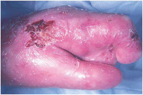
– Mitten deformities of hands and feet develop with digital fusion (Figure 85.6).
– Teeth often are carious; delayed eruption may be noted.
– Microstomia develops from scarring.
– Other complications include difficulty swallowing (esophageal scarring), chronic anemia, growth failure, conjunctival scarring, and predisposition to squamous cell carcinoma.
Figure 85.1. Epidermolysis Bullosa Simplex (Localized or Weber-Cockayne Type). This Patient Has a Bulla Involving the Great Toe and a Healing Bulla on the Ball of the Foot.
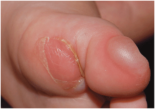
Figure 85.2. Numerous Bullae and Erosions in a Patient with Junctional Epidermolysis Bullosa, Generalized Severe (Herlitz Type).
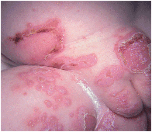
Figure 85.3. Denudation of the Lower Leg and Foot in a Newborn with Junctional Epidermolysis Bullosa with Pyloric Atresia. She Died from Overwhelming Infection Shortly after Birth.
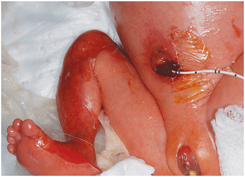
Figure 85.4. Multiple Milia with Scarring over the Dorsal Aspect of the Hand and Fingers of a 1-Year-Old with Dominant Dystrophic Epidermolysis Bullosa.
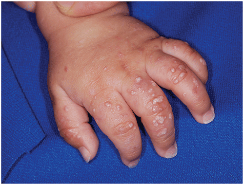
Figure 85.5. This Young Adult with Recessive Dystrophic Epidermolysis Bullosa Has Widespread Bullae and Erosions that Heal with Scarring.
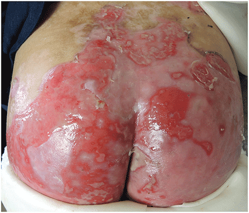
Figure 85.6. Mitten Deformity of the Hand of a Patient with Recessive Dystrophic Epidermolysis Bullosa.
Look-alikes
| Disorder | Differentiating Features |
|---|
| Bullous congenital ichthyosiform erythroderma | Blisters may be present soon after birth, similar to epidermolysis bullosa. Thickened areas of skin with ridging often present during infancy or develop with time. Eventuates into an ichthyosis disorder (epidermolytic hyperkeratosis), with less propensity toward blistering.
|
| Incontinentia pigmenti | Small vesicles occur in clusters. Blisters are arranged in a linear or whorled pattern, along Blaschko lines. Subsequent to blister stage, skin lesions appear verrucous or hyperpigmented. Most patients are female (X-linked dominant). Blisters not trauma induced.
|
| Bullous impetigo | Does not usually present as a recurrent or chronic condition. Involvement more focal. Mucous membranes not involved. Blisters rupture easily, leaving superficial erosions with peripheral collarettes of scale. Blisters not trauma induced.
|
| Herpes simplex virus infection | |
| Bullous pemphigoid | Urticarial plaques present in addition to tense blisters. Blisters not trauma induced. Pruritus common with early lesions. Direct fluorescence and immunoblotting studies will help confirm diagnosis.
|
| Dermatitis herpetiformis | Usually presents as tiny vesicles and erosions. Most often clustered on elbows, knees, shoulders, sacrum, and buttocks. Blisters not trauma induced. Pruritus is intense. May be associated with gluten sensitivity.
|
| Erythema multiforme major | Typical target lesions may be present. Only occasionally bullous, and bullae are not trauma induced. Oral mucous membrane erosions common. Palms and soles usually involved. History of herpes simplex virus infection or drug ingestion may be present.
|
| Acquired epidermolysis bullosa | Acquired autoimmune blistering disease, not genetic. Direct fluorescence and immunoblotting studies will help confirm diagnosis.
|
| Linear IgA dermatosis | Acquired autoimmune blistering disorder, not genetic. “Cluster of jewels” pattern (annular grouping of bullae) often noted. Blisters not trauma induced. Mucosal involvement not as extensive as in epidermolysis bullosa.
|
▶Molecular genetic testing is recommended to confirm the diagnosis and aid in prognosis and decision-making.
▶Skin biopsy for immunomapping, when available, may provide for more prompt diagnosis.
▶Electron microscopy may be helpful when genetic testing or immunomapping is not available or does not provide conclusive results, but this is rarely used in the current era.
▶Prenatal genetic testing is possible and should be considered when applicable.
▶Treatment is palliative and supportive.
▶Avoidance of trauma, treatment of infections, pain control, and nutritional counseling are all vital.
▶Bullae may be drained with sterile needle and syringe for pain control.
▶Antibiotic ointment and protective dressings can be applied to areas of open or blistered skin to promote healing and prevent secondary infection.
▶Patient and family education, psychologic support, and referral to support group organizations are important.
▶Multidisciplinary treatment teams are important to decrease morbidities associated with EB.
▶Care teams may include representation from primary care and pediatrics, dermatology, nursing, plastic surgery, ophthalmology, gastroenterology, general surgery, hematology, dentistry, genetics, and nutrition.
▶Growth failure is treated with aggressive nutritional rehabilitation; gastrostomy tube placement may be required for infants with severe forms of EB.
▶Esophageal involvement with dysphagia may require dietary modifications or dilatation procedures.
▶Mitten deformities of the hand require physical therapy and surgical intervention (degloving procedures).
▶Prognosis depends on the subtype of EB.
▶Children with most forms of EBS, non-Herlitz JEB, and dominant DEB tend to have a fairly good prognosis.
▶EBS, generalized severe (Dowling-Meara) subtype, may be severe during infancy and is occasionally fatal.
▶Children with Herlitz JEB and JEB with pyloric atresia have a poor prognosis.
▶Patients with recessive DEB have a chronic course marked by complications and diminished quality of life. Squamous cell carcinoma, if it occurs, is usually rapidly progressive and invasive, leading to death in most of these patients.
▶Patients with possible EB should be referred to an experienced dermatologist for confirmation of the diagnosis and coordination of multidisciplinary care.
▶Because of the increased risk of cutaneous squamous cell carcinoma, biopsy should be performed in any suspicious lesion in patients with recessive DEB.
▶American Academy of Dermatology: Epidermolysis bullosa: overview.
https://www.aad.org/public/diseases/a-z/epidermolysis-bullosa-overview
▶Dystrophic Epidermolysis Bullosa Research Association (debra) of America: Provides information, support, and resources for patients who have epidermolysis bullosa and their families.
www.debra.org
▶Dystrophic Epidermolysis Bullosa Research Association (debra) UK: Located in the United Kingdom, this organization provides information for patients who have epidermolysis bullosa and their families.
www.debra.org.uk
▶Epidermolysis Bullosa Medical Research Foundation: Dedicated to supporting research in EB: Provides information for patients and families.
https://www.ebmrf.org