▶Periorificial dermatitis (formerly known as perioral dermatitis) is an acneiform disorder of facial skin initially described in older teenagers and young adult women but also seen in younger children.
▶Some consider this a pediatric form of acne rosacea, displaying a combination of acneiform papules and pustules along with varying degrees of erythema in a periorificial distribution.
▶A granulomatous juvenile variant of classic periorificial dermatitis exists. It is sometimes referred to as granulomatous periorificial dermatitis.
▶The cause is unknown, but it has been associated with chronic application of topical corticosteroids (or use of steroids in other forms, including inhaled), as well as bubble gum, oils, greases, and fluoridated toothpastes.
▶Both male and female patients can be affected.
▶Erythematous to flesh-colored monomorphous papules and papulopustules distributed around the mouth, with a narrow zone of sparing around the vermilion border (Figures 9.1 and 9.2).
▶Scaling may be present.
▶Comedones are absent.
▶Other periorificial areas are commonly affected, including the peri-nasal and periorbital regions. There may be a history of recurrent chalazion and hordeolum (sty).
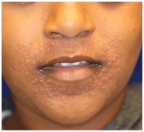
▶The granulomatous variant presents in a similar fashion but with prominence of translucent, pink to flesh-colored or tan papules (Figure 9.3) and often a history of prior corticosteroid application.
Figure 9.1. Periorificial Dermatitis. Erythematous Papules and Papulopustules around the Nares and Mouth, with a Rim of Sparing around the Vermilion Border.
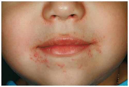
Figure 9.2. Periorificial Dermatitis. Erythematous Fine Papules Previously Treated with Topical Corticosteroids around the Nares and Mouth in a Young Child.
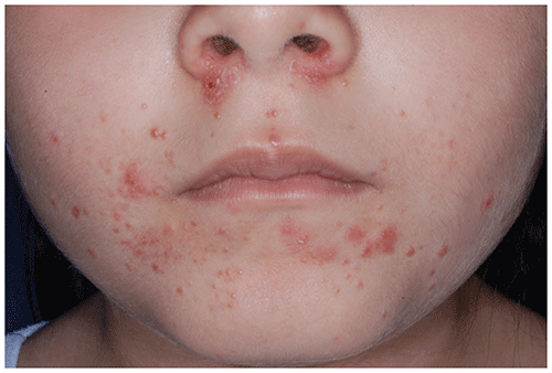
Figure 9.3. Granulomatous Periorificial Dermatitis. Flesh-Colored to Pink, Translucent Papules in the Perioral and Perinasal Locations, with Some Lower Periorbital Involvement, in a Child.
Look-alikes
| Disorder | Differentiating Features |
|---|
| Atopic dermatitis | Less papular and typically more scaly. Other areas of body usually affected. Pruritus usually present.
|
| Acne vulgaris | Distribution of early lesions more commonly in the T-zone (ie, forehead, nose, and chin). Comedones present. Onset rarely occurs between 1 and 7 years of age.
|
| Allergic contact dermatitis | Less papular and may be more geometric in distribution. History of exposure to antigen may be present. Pruritus usually present and significant.
|
| Irritant contact dermatitis (eg, caused by lip licking or pacifier) | Less papular and may be more geometric in distribution. History of lip licking or pacifier use present. Involved areas often have sharp geometric borders. Vermilion border is typically involved.
|
| Flat warts | Small, skin-colored to tan, flat-topped papules may coalesce into plaques. Erythema typically absent. May be present elsewhere on the body. Koebnerization (ie, distribution of lesions in a linear fashion after skin trauma or scratching) may be evident.
|
| Sarcoidosis | When limited to face, may be difficult to distinguish from periorificial dermatitis. Papules often reddish brown. Often in other locations (eg, neck, upper trunk, extremities).
|
| Benign cephalic histiocytosis | Usually occurs in children 3 years or younger. Papules may be erythematous, but often yellowish brown, and may simulate flat warts. Typically not periorificial in distribution.
|
▶The diagnosis is made clinically on the basis of lesion morphology and characteristic distribution.
▶Skin biopsy may be useful in questionable cases but is rarely necessary.
▶Mild to moderate cases: topical calcineurin inhibitors (eg, pimecrolimus cream, tacrolimus ointment) or topical antibiotics, most commonly metronidazole or erythromycin, applied once to twice daily. Topical sulfacetamide with or without sulfur may also be useful.
▶Severe cases: oral antibiotic therapy (eg, with erythromycin, azithromycin; in patients older than 8 years, doxycycline or minocycline) for a minimum of 6 to 8 weeks with gradual tapering to avoid rebound flaring.
▶Treatment consideration: Topical corticosteroids lead to initial improvement in periorificial dermatitis, but rapid flaring is seen when they are discontinued. If the patient’s condition has been treated with this agent, consider tapering the potency of the topical steroid gradually over weeks; another option is to substitute the topical steroid with a topical calcineurin inhibitor as monotherapy or concurrently with a systemic antibiotic (if more severe).
▶The condition improves slowly (often requires 4–12 weeks) but steadily with appropriate therapy.
▶Treatments should be used until clearing has occurred, with gradual tapering to prevent rebound.
▶Postinflammatory hyperpigmentation or hypopigmentation may be seen and generally resolves over several months.
▶Consider referral if the diagnosis is in question or the condition has not responded to appropriate therapy.