▶Caused by human parvovirus B19.
▶Usually affects children between 4 and 10 years of age.
▶Most common in the winter and spring, with endemic peaks every 6 to 9 years.
▶Transmission is via respiratory droplets, blood and blood products, or vertically from mother to fetus.
▶Incubation period is 4 to 14 days.
▶Clearance of viremia precedes appearance of the erythema infectiosum rash by several days; thus, patients who have the skin eruption are not considered contagious.
▶Up to 50% of infections may be subclinical.
▶During the viremic stage, patients may develop prodromal symptoms of fever, malaise, myalgias, pharyngitis, and headache.
▶After the viremic phase, the first stage of erythema infectiosum reveals the classic finding of a slapped cheek appearance with bright red erythema on the cheeks, typically sparing the perioral region (Figures 14.1 and 14.2).
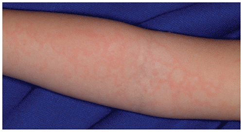
▶The second stage appears 1 to 4 days after the facial rash appears and is characterized as erythematous patches, papules, and plaques that partially clear, leaving a lacy, reticular pattern of erythema, especially on the flexor surfaces of the arms. This phase of the exanthem may wax and wane over the next 1 to 4 weeks (Figure 14.3).
▶Pruritus is sometimes prominent.
▶After the exanthem fades, it is commonly reactivated for several weeks to months by physical factors, including sunlight, physical activity, or hot baths.
▶Arthralgia and arthritis may be the most common manifestation of parvovirus B19 infection in adolescents and adults, especially if female. However, this is less frequent in younger children. The joint symptoms are typically brief in duration, preferentially affecting larger joints, and may be pauciarticular or polyarticular. Rarely, the arthralgia may persist for months to years.
▶Human parvovirus B19 exhibits tropism for erythroid progenitor cells, and individuals with predisposing hematologic conditions resulting in a shortened red blood cell half-life (eg, sickle cell disease, spherocytosis, thalassemia) are at risk for aplastic crises. These crises occur before and in the early periods of the exanthem.
▶Pregnant women who are susceptible and become infected with human parvovirus B19 during the first half of their pregnancy may transmit the infection to their developing fetus, with subsequent risk of fetal anemia, nonimmune fetal hydrops, and fetal death in 2% to 6% of cases.
Figure 14.1. The First Stage of Erythema Infectiosum in a Child with Skin of Color. There are Erythematous Patches on the Cheeks. From Redbook Visual Library. Courtesy of H. Cody Meissner, Md, Faap.
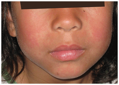
Figure 14.2. The First Stage of Erythema Infectiosum Exhibits Erythematous Cheeks (Ie, a Slapped Cheek Appearance).
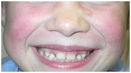
Figure 14.3. The Second Stage of Erythema Infectiosum Produces an Erythematous Lacy, Reticulated Exanthem on the Extremities.
Look-alikes
| Disorder | Differentiating Features |
|---|
| Exanthematous drug eruption | History of drug exposure elicited. Slapped cheek appearance absent. Typically presents with red macules and papules.
|
| Nonspecific viral exanthem | Fever or other symptoms may be present. Slapped cheek appearance absent. Typically presents with red macules and papules.
|
| Livedo reticularis | |
| Exanthem of juvenile idiopathic arthritis | Clinical features of juvenile idiopathic arthritis present. Slapped cheek appearance absent. Exanthem most apparent during febrile periods.
|
| Scarlet fever | Circumoral pallor may give slapped cheek appearance. Generalized, sandpaper-like eruption. Pharyngitis and lymphadenopathy usually present.
|
| Urticaria | Acute onset of pruritic and edematous papules, plaques, wheals. Distribution usually generalized. Dermatographism may be present. Lesions last less than 24 hours and migrate to other areas.
|
▶The diagnosis is most often made clinically on the basis of characteristic findings.
▶Serological detection of immunoglobulin M directed against human parvovirus B19 can confirm the diagnosis when obtained within 30 days of illness onset.
▶No specific treatment is indicated.
▶Children with the characteristic rash can return to school or child care, as they are no longer considered contagious.
▶Nonsteroidal anti-inflammatory drugs may be used for arthritis.
▶Hospitalization and red blood cell transfusion may be required in children with transient aplastic crises.
▶Erythema infectiosum typically resolves without sequelae.
▶Patients with parvovirus B19 infection who are immunodeficient may develop chronic bone marrow suppression, and intravenous immunoglobulin therapy has been used in this setting.
▶Pregnant women who have been exposed should be advised to contact their obstetric health professional to discuss potential risks and be offered serological testing. If acute infection is confirmed, serial fetal ultrasonography should be considered to monitor for fetal hydrops, congestive heart failure, and intrauterine growth restriction.
▶Referral may be indicated if atypical features are present or the diagnosis is in question.
▶Pregnant women exposed to or infected with human parvovirus B19 should consult their obstetric health professional (as discussed previously).
▶Patients who are immunocompromised and patients with predisposing blood disorders who are exposed to human parvovirus B19 should be monitored for signs and symptoms of aplastic crises.