▶Ecthyma is a deep and ulcerative pyoderma (a deep cutaneous infection) that is most prevalent in tropical climates.
▶The most common causative organisms are group A β-hemolytic streptococci (GABHS) and Staphylococcus aureus.
▶Although the lesions may initially seem to be impetigo, the organisms progress to invade beyond the epidermis and deeper into the dermis.
▶Ecthyma can develop at sites of previous skin disorders, such as insect bites or scabies.
▶The extremities and buttocks are most often involved.
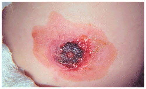
▶Ecthyma lesions may be vesicopustules or crusted erosions; usually there is surrounding erythema. Often lesions will progress to become necrotic in appearance, with deep punched-out ulcers (Figure 25.1). The ulcer can be saucer shaped with a raised surrounding border.
▶Lesions are often crusted, and removal of the crust reveals a deeper ulcer.
▶Lesions are painful and often multifocal; they heal slowly over a few weeks, often with scar formation.
Figure 25.1. Ecthyma Lesion with Central Necrotic Crust.
Look-alikes
| Disorder | Differentiating Features |
|---|
| Impetigo | Non-bullous |
BullousFragile bullae that rupture rapidly leaving round superficial erosions. Periphery of lesions may exhibit a collarette of scale, which is the remnant of the bulla roof.
|
| Brown recluse spider bite | Timing of lesion appearance corresponds with exposure to spider. Usually becomes painful after a few hours. Usually a single location rather than a multifocal process. Initial lesion noted to have central hemorrhagic area with surrounding edema and erythema; may produce the “red, white, and blue sign” (ie, rings of color surrounding the lesion). Rapidly progresses, resulting in large necrotic plaque, commonly with eschar formation.
|
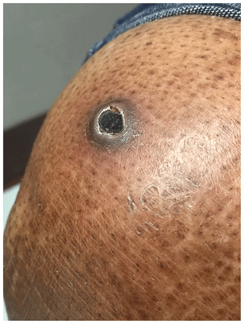
| Ecthyma gangrenosum | Localized septic vasculitis usually associated with Pseudomonas aeruginosa bacteremia. Lesions initially present as hemorrhagic papules. Subsequently progresses into deep ulcer with necrosis (Figure 25.2) and, occasionally, eschar formation. Often accompanied by high fever, myalgias. Most children who are affected are immunosuppressed.
|
| Cutaneous anthrax | Spores enter through a cut or an abrasion. Initial lesion is a painless, pruritic papule that subsequently develops a central clear bulla. When bulla ruptures, necrosis and an ulcer develop, surrounded by massive edema and multiple smaller lesions.
|
| Vasculitis | Many vasculitic processes can present with hemorrhagic papules with necrosis, including hypersensitivity vasculitis, Henoch-Schönlein purpura, and polyarteritis nodosa. Associated symptoms (eg, fever) and other signs (eg, hematuria, hematochezia, arthritis) may assist in diagnosis and help to differentiate these disorders from ecthyma.
|
| Cigarette burns | |
Figure 25.2. Ecthyma Gangrenosum Lesion in a Patient with Skin of Color. Note the Central Crusted Ulceration with Surrounding Faintly Visible Erythema. Reproduced with Permission from Khoo T, Ford F, Lobo Z, Psevdos G. One Thing after Another: Ecthyma Gangrenosum. Am J Med. 2018;131(5):510–511.
▶The clinical findings usually lead to the correct diagnosis.
▶Bacterial culture often reveals GABHS.
▶Skin biopsy (usually unnecessary) shows an intense polymorphonuclear infiltrate and organisms on tissue Gram stain.
▶Systemic antibiotic therapy is the treatment of choice.
▶The chosen antibiotic should have activity against GABHS and S aureus (because the latter is an occasional cause or may be present as a secondary infectious agent). Failure of therapy indicates the need to review culture and sensitivity results for presence of methicillin-resistant S aureus or to reconsider the diagnosis.
▶Because of the penetration into the dermis, ecthyma lesions often heal with permanent scarring.
▶Rapid healing usually occurs with appropriate antibiotic therapy.
▶Consider referral to a dermatologist for patients who have severe or extensive disease or in whom standard treatment does not work.