▶Hamartoma of sebaceous and apocrine glands and epidermal elements present in 0.3% of newborns.
▶Usually appears as an isolated finding; rarely associated with neurologic, ocular, or skeletal variations (ie, epidermal nevus or Schimmelpenning syndrome).
▶Usually presents at birth as a solitary, well-circumscribed round or oval plaque.
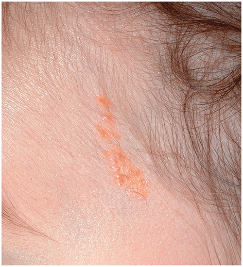
▶Typically located on the scalp, where it is associated with alopecia (Figures 107.1 and 107.2), or face (Figure 107.3).
▶Lesions are yellow, yellowish brown, orange, or pink and have a velvety or verrucous (wartlike) texture. They are often linear or curvilinear.
▶At puberty, androgenic stimulation causes lesions to become more elevated and verrucous, with development of a rough surface.
Figure 107.1. Nevus Sebaceus. Yellowish Tan Hairless Plaque Located on the Scalp.
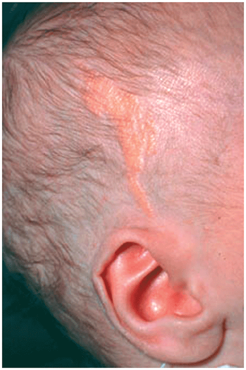
Figure 107.2. Large Nevus Sebaceus on the Scalp of a Young Child with Skin of Color. Note the Hairless Pink to Brown Plaque. These Lesions May Not Always Appear Yellowish Orange in Color, Especially in Those with Darker Skin Tones. Reproduced with Permission from Dermnet Nz.
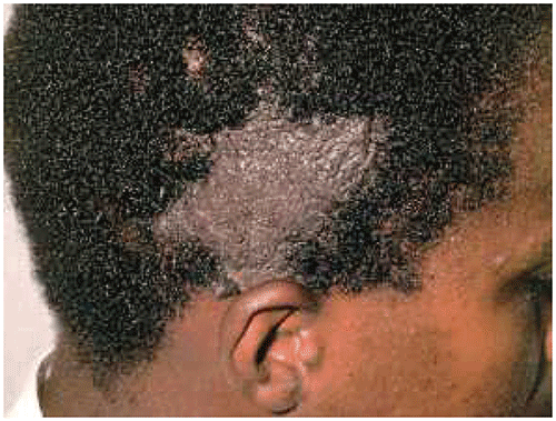
Figure 107.3. A Linear Nevus Sebaceus Located on the Face.
Look-alikes
| Disorder | Differentiating Features |
|---|
| Aplasia cutis congenita | |
| Epidermal nevus | |
| Juvenile xanthogranuloma | |
▶No treatment is required during infancy or childhood.
▶Because there is a small risk of developing basal cell carcinoma (and, rarely, other cutaneous malignancy) within the nevus after puberty, some advise elective excision before that time. Clinical observation is a reasonable alternative for lesions that are not psychosocially concerning and that remain stable. If secondary neoplasms develop within an existing nevus sebaceus, surgical excision with histopathologic analysis is strongly recommended.
▶Most sebaceous nevi exhibit a benign course, although there is a small chance of secondary neoplasms (including malignant transformation), as discussed.
▶Changes in a nevus sebaceus (eg, development of a nodule) should prompt referral to a dermatologist.
▶If concern exists about epidermal nevus syndrome (eg, the nevus sebaceus is extensive or linear and associated with developmental delay, seizures, or ophthalmologic variations [eg, coloboma of the eyelid]), consultations with a pediatric dermatologist, neurologist, medical geneticist, and ophthalmologist should be sought, as indicated.