▶Congenital melanocytic nevi (CMN) are found at birth in about 1% of newborns.
▶CMN have an increased risk of malignant transformation.
■The risk of malignant transformation in small (<1.5 cm) congenital nevi is quite low and considered by most experts to be no greater than the baseline risk of malignant melanoma in the general population.
■The risk of malignant transformation in medium (1.5–20 cm), large (>20–40 cm), or “giant” (>40 cm) congenital nevi is higher than the baseline risk in the general population but difficult to quantify; some studies of giant congenital nevi have estimated this risk to be as high as 10% to 15%.
■Importantly, a substantial number of the melanomas arising within a large or giant congenital melanocytic nevus may occur in the central nervous system, rather than the skin.
▶CMN usually are present at birth but may appear during the first 6 months after birth.
▶Most lesions are small (<1.5 cm in diameter). Large CMN occur in about 1 in 20,000 newborns and giant CMN in around 1 in 500,000 newborns.
■Congenital nevi are typically larger than acquired nevi and have significant hair growth within them (Figure 71.1).
■They are usually slightly elevated and have surface texture changes (Figure 71.2).
▶Occasionally, lesions are larger, measuring 20 cm or more (Figure 71.3).
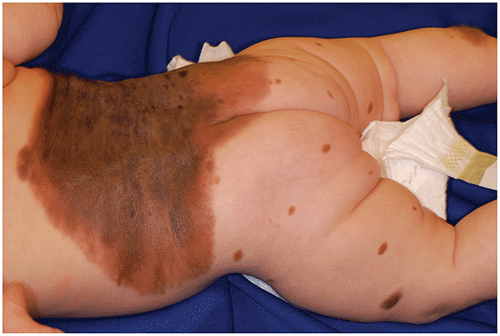
▶Large and giant CMN are often accompanied by satellite lesions (Figure 71.4).
Figure 71.1. Congenital Melanocytic Nevus with Superimposed Hypertrichosis.
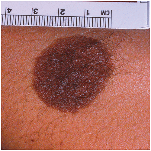
Figure 71.2. Congenital Melanocytic Nevus Demonstrating Surface Textural Change.

Figure 71.3. A Giant Congenital Melanocytic Nevus Involving the Entire Posterior Trunk.

Figure 71.4. A Large Congenital Melanocytic Nevus Involving the Posterior Trunk. There are Multiple Satellite Nevi Present as Well.
Look-alikes
| Disorder | Differentiating Features |
|---|
| Ephelides | Small, hyperpigmented macules located in sun-exposed areas such as the face, upper chest, and back. Ephelides become darker after sun exposure and may lighten during times of less sun exposure. Unlike congenital melanocytic nevi (CMN), have no change in surface texture, hypertrichosis, or malignancy potential.
|
| Lentigines | Small, hyperpigmented macules not limited to sun-exposed areas. Unlike CMN, have no change in surface texture, hypertrichosis, or malignancy potential.
|
| Café au lait macules | |
| Becker melanosis (Becker nevus) | |
| Plexiform neurofibroma | May present with “bag of worms” consistency on palpation (representing enlarged nerve roots). May be associated with pain, atrophy, muscle loss. Risk of malignant degeneration into malignant peripheral nerve sheath tumor. Associated with neurofibromatosis type 1 (considered one of the diagnostic criteria) (see also Chapter 89).
|
▶Small CMN that are asymptomatic and not changing may be observed or excised at puberty (malignant change before puberty is extraordinarily rare).
▶Infants who have larger lesions should be referred to a plastic surgeon for discussion of possible excision and to a dermatologist for clinical surveillance (if not excised).
▶Infants with extensive CMN on the head or overlying the midline of the back, and especially in those with large numbers of satellite nevi, are at risk of central nervous system involvement (ie, neurocutaneous melanosis). Magnetic resonance imaging of the brain and spinal cord should be performed.
▶Medium, large, and giant CMN are at increased risk of melanoma development and should be considered for excision, when feasible.
▶Close clinical follow-up for atypical progression (eg, ulceration, new nodular components, bleeding, color change) with biopsy of suspicious areas is vital for patients who have not had such larger congenital nevi excised.
▶Melanoma is the malignant neoplasm of melanocytes that may arise de novo or from preexisting nevi. Consider the possibility of melanoma when a CMN exhibits any of the following ABCDE criteria:
■Asymmetry.
■Border irregularity.
■Color variation (especially red, blue, black).
■Diameter larger than about 6 mm (but this criterion is less useful for CMN, as they are often larger than this very early after birth).
■Evolving lesion that is changing quickly.
▶Recently, it has been suggested that applying these traditional ABCDE criteria in addition to modified ABCD criteria may facilitate earlier detection of melanoma in children. The modified ABCD criteria (and explanation) are as follows:
■Amelanotic (as pediatric melanoma may be nonpigmented).
■Bleeding, bump (bleeding lesions; lumps or bumps [ie, papulonodules or papules]).
■Color uniformity (as the color may be consistent and uniform throughout the lesion).
■De novo, any diameter (new papular lesions; lesions may be <6 mm).
▶Patients with CMN that are medium, large, or giant in size or which show atypical features or any symptoms (eg, rapid evolution, nonhealing sores or bleeding) should be referred for prompt dermatologic evaluation.