▶Herpes simplex virus (HSV) 1 (HSV-1) and HSV-2 are 2 members of the Herpesviridae family of viruses that also includes varicella-zoster virus (VZV); Epstein-Barr virus; cytomegalovirus; and human herpesviruses 6, 7, and 8.
▶Primary HSV infection is generally a childhood disease involving the mouth (herpetic gingivostomatitis), lips, or eyes.
■Serological evidence of HSV-1 infection increases steadily with age, and 18% to 35% of children are estimated to have infection by 5 years of age.
■In many cases, acquisition of HSV infection does not cause symptoms.
■If acquisition of infection is accompanied by clinical disease, it is characterized as primary clinical disease. Such syndromes (eg, primary HSV gingivostomatitis) are typically moderate to severe illnesses accompanied by fever, lymphadenopathy, constitutional symptoms, and more severe or prolonged cutaneous or mucocutaneous disease.
▶HSV infections are characterized by the phenomenon of latency. After initial infection, individuals are prone to subsequent recurrences of localized cutaneous disease at the site of initial infection resulting from reactivation of latent virus in regional sensory or autonomic nerve ganglia. Recurrent HSV is generally a more limited clinical syndrome than primary HSV.
▶Asymptomatic acquisition of primary HSV infection may subsequently lead to clinically recognized disease, often with features of recurrent HSV (ie, milder disease).
▶HSV infections are typically transmitted by direct contact with skin lesions or infectious mucous membrane secretions.
▶HSV-2 infection is most commonly acquired by sexual contact; however, HSV-1 prevalence in anogenital infections has been increasing.
■Genital HSV-2 infection in prepubertal children should raise suspicion of child abuse.
■HSV-2 is the most common cause of neonatal HSV infection globally. The yearly incidence of neonatal HSV has increased in the United States, occurring in as many as 5 per 10,000 deliveries in 2015.
▶Neonatal HSV (Figure 10.1) most commonly occurs as a result of transmission during passage through the birth canal, less often via ascending infection. It is rarely caused by postnatal transmission from a caregiver.
■Neonates born to mothers who have a primary genital infection are at the highest risk (25%–60%) of becoming infected.
■Risk of transmission to newborns by mothers shedding HSV as the result of a reactivated infection is significantly lower (2%).
■Approximately two-thirds of neonates with disseminated or central nervous system disease have skin lesions, but these lesions may not be present at onset of symptoms.
Figure 10.1. Clustered Vesicles on an Erythematous Base in an Infant with Neonatal Herpes Simplex Virus Infection.
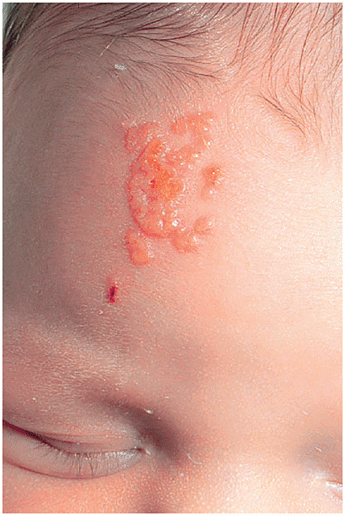
▶Grouped 1- to 2-mm vesicles on an erythematous base are the classic lesions of HSV skin infection (Figure 10.1). In darker skin types, the erythematous base may appear more violaceous (Figure 10.2).
▶Skin vesicles evolve to form pustules, erosions, and crusts.
▶Mucosal vesicles in areas of friction (eg, mouth, vulvovaginal area, anorectal area) are rapidly unroofed and form small ulcers.
▶Coalescent vesicles or erosions may appear as larger bullae or superficial ulcerations.
▶Lesions may occur on any cutaneous or mucocutaneous site but are most common on the face.
▶Skin lesions are sometimes pruritic but typically painful. Primary infection syndromes may cause severe pain.
▶Regional lymphadenopathy is common, particularly with primary HSV infection.
▶Recurrent HSV infection is often preceded by a characteristic brief prodrome of itching or dysesthesia at the site of impending recurrence.
▶Distinctive regional distributions of HSV infection are recognized as the following clinical syndromes:
■Primary HSV gingivostomatitis: Children who are affected develop ulcers on the buccal mucosae, tongue, gingivae, and perioral skin (Figure 10.3). These findings may be accompanied by fever, lymphadenopathy, and constitutional symptoms.
■Herpes labialis (cold sore): Lesions occur on the lips, most often at the vermilion border; most common type of recurrent herpes infections overall (Figure 10.4).
■Herpetic whitlow: Deep-seated painful vesicles on the distal portion of the finger; may be primary or recurrent HSV infection (Figure 10.5).
■Genital HSV infection: In addition to mucocutaneous symptoms, secondary symptoms may include fever, swelling, and dysuria.
■Ocular HSV infection: Corneal involvement may cause keratoconjunctivitis, which may be associated with pain, photophobia, and eye discharge.
■Herpes gladiatorum: Typically seen in participants of contact sports, and characteristic lesions may affect the head, neck, and upper extremities. Rarely, the cutaneous findings may lack vesicles.
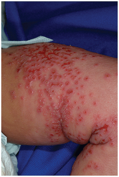
■Eczema herpeticum: severe, widespread HSV infection in an individual with preexisting generalized skin disease, most often atopic dermatitis (Figure 10.6). Patients may present with fever, malaise, and numerous monomorphic vesicles and heme-crusted, punched-out erosions in areas of active underlying skin disease.
■Zosteriform herpes simplex: manifests as recurrent shingles.
Figure 10.2. Grouped Vesicles in Herpes Simplex Virus Infection with Central Heme Crust and Mild Underlying Violaceous Color Changes in a Patient with Skin of Color.
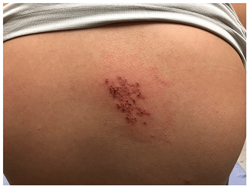
Figure 10.3. Grouped Vesicles and Ulcers Affecting the Perioral Skin are Observed in Herpes Simplex Virus Gingivostomatitis. Note that the Periphery of Grouped Vesicles May Form a Scalloped Appearance.
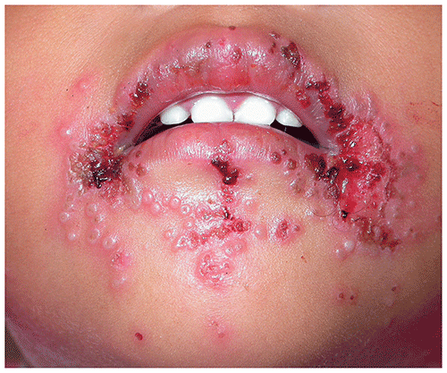
Figure 10.4. Herpes Labialis (Cold Sore). Vesicles Occur on the Lips, Most Often at the Vermilion Border.
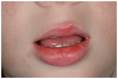
Figure 10.5. Deep-Seated Vesicles with Surrounding Erythema and Swelling Located on the Finger are Characteristic of Herpetic Whitlow.
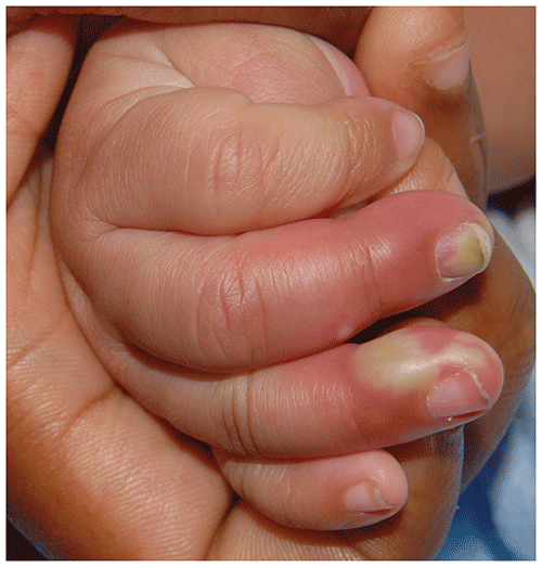
Figure 10.6. Eczema Herpeticum is Characterized by Numerous Vesicles and Monomorphous Erosions with a Punched-Out Appearance, Typically in Areas of Active Dermatitis.
Look-alikes
| Disorder | Differentiating Features |
|---|
| Herpes zoster | |
| Allergic contact dermatitis | |
| Hand, foot, and mouth disease | Individual (not grouped) vesicles. Palm and sole involvement is a prominent feature. Vesicles often oval. With severe disease, larger bullae may be present. In “eczema coxsackium,” lesions may predominate in areas prone to atopic dermatitis, may be numerous, and may be clustered (ie, closely simulates eczema herpeticum).
|
| Herpangina | Vesicles and erosions primarily involve the palate, uvula, and tonsillar pillars. No involvement of surrounding perioral skin or lips.
|
| Aphthous stomatitis (canker sores) | Single or multiple (usually ≤3) discrete, shallow ulcers, 3 to 6 mm in size, affecting the oral mucosa. Lesions have grayish white membrane and sharp, slightly raised, red borders.
|
| Bullous impetigo | Flaccid bullae or round, superficial erosions with a rim of surrounding scale. No deep-seated vesicles as seen in HSV infection. Culture grows Staphylococcus aureus.
|
| Blistering distal dactylitis | May be difficult to distinguish clinically from herpetic whitlow. Solitary bulla, whereas the lesions of HSV infection often are smaller vesicles. Culture typically positive for Streptococcus pyogenes, less often S aureus.
|
| Thermal burn | |
| Bullous mastocytoma | Peau d’orange appearance of surface, often with hyperpigmentation. History of localized blistering, which may become less likely after infancy. Positive Darier sign (urtication after firm stroking of lesion).
|
▶The diagnosis usually is made clinically on the basis of the classic clinical morphology and distribution of lesions, especially when supported by history of recurrence.
▶Laboratory investigations can be useful when the diagnosis is uncertain.
■Viral culture is a reliable method for confirming the diagnosis and is considered the criterion standard, although, in many settings, it has been replaced by polymerase chain reaction testing.
■Polymerase chain reaction testing, when available, is a highly sensitive and specific diagnostic test.
■Direct immunofluorescence examination of lesional swabs offers rapid diagnosis and can distinguish HSV and VZV infection with high sensitivity, but such testing may not be available in many office settings.
■Serological studies are less useful clinically.
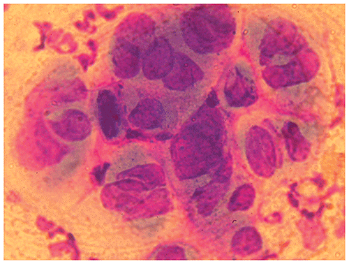
■Tzanck test of a fresh vesicle can provide rapid information, but utility of the test is limited by experience or expertise of the clinician and hampered by suboptimal sensitivity and specificity (Figure 10.7). A positive Tzanck test result cannot distinguish between HSV and VZV infection.
Figure 10.7. Tzanck Test in Herpes Simplex Virus Infection; Multinucleated Giant Cells are Present.
▶Supportive therapy suffices in most cases, using simple cleansing and comfort measures, astringent gels, soothing moisturizers, or topical antibiotic ointments (to prevent secondary bacterial infection).
▶Oral analgesics (eg, viscous lidocaine solution, benzocaine lozenges, compounded mouthwashes) may be useful for associated pain.
▶Oral antiviral therapy is indicated for severe disease, frequently recurrent disease, and individuals who are immunosuppressed. Oral acyclovir, valacyclovir, or famciclovir may be used, depending on the patient’s age. Treatment is more effective when started earlier (eg, in the first 48 hours) in the outbreak.
▶Severe primary infections, infections in those who are immunocompromised, recurrences associated with erythema multiforme, and eczema herpeticum should be treated systemically (oral or intravenous, depending on severity and host risk factors); some of these patients may merit inpatient hospitalization.
▶Neonatal HSV infection requires parenteral acyclovir therapy.
▶The decision to treat less severe or recurrent outbreaks is based on the frequency and severity of the lesions and level of distress to the patient or family.
▶Current treatment guidelines are summarized in the current edition of AAP Red Book®: Report of the Committee on Infectious Diseases (https://redbook.solutions.aap.org).
▶In most individuals who are immunocompetent, HSV infections beyond the neonatal period are mild and self-limited and have an excellent prognosis.
▶Circumstances in which disease may be more severe and require more aggressive therapy are discussed in the next section.
▶Newborns and infants 6 weeks or younger who have evidence of HSV infection should be evaluated immediately and treated with intravenous acyclovir. Consultation with a pediatric infectious disease specialist is desirable.
▶Widespread lesions over eczematous skin (ie, eczema herpeticum) or infection in a child who is immunocompromised can be severe, often requiring hospitalization and treatment with intravenous acyclovir.
▶Widespread oral lesions (ie, HSV gingivostomatitis) with resulting mouth pain and dehydration require hydration, antiviral therapy, and analgesia.
▶Patients who develop erythema multiforme or Stevens-Johnson syndrome after HSV infection should be referred to a dermatologist and often require suppressive antiviral therapy in an effort to prevent recurrences.
▶Involvement in or around the eye should be immediately evaluated by an ophthalmologist.
▶Evidence of central nervous system involvement (eg, seizures, behavioral changes, lethargy) warrants emergent evaluation.
▶Sexual abuse should be suspected in children with anogenital herpes infection if there is no clear history of autoinoculation as the source of infection.