▶Rare inflammatory skin disorder of unknown etiology.
■CARD14 pathogenetic variants identified in a subset of patients with hereditary autosomal dominant form.
▶Affects patients of all ages with 2 peaks of onset: 1 in the first decade after birth and another in adulthood. Although most pediatric patients develop symptoms in the teenage years, the disorder can also be seen during the first couple years of life.
▶Most common differential diagnosis: psoriasis.
▶Clinical presentation can be further subdivided into adult onset (classic or atypical) and childhood onset (classic juvenile, circumscribed juvenile, and atypical juvenile). The features of childhood onset pityriasis rubra pilaris (PRP) are summarized in Table 53.1.
▶Circumscribed juvenile PRP is the most common subtype seen in children.
Table 53.1. Features of Childhood Onset Pityriasis Rubra Pilaris (PRP)
| Juvenile PRP Type | Frequency | Clinical Features |
|---|
| Classic juvenile (type III PRP) | 14%–35% | Generalized pattern. Follicular keratotic papules. Patients with extensive disease may demonstrate islands of uninvolved skin. Palm and sole involvement common.
|
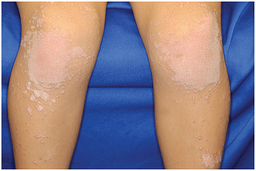
| Circumscribed juvenile (type IV PRP) | Most common subtype in children | Focal hyperkeratotic papules and plaques on the extensor surfaces of elbows, knees (Figure 53.3). Palmoplantar lesions common.
|
| Atypical juvenile (type V PRP) | Rare; may be familial and can have younger age of onset | |
▶Hyperkeratotic papules and plaques, often surrounding the hair follicles and demonstrating a salmon-colored hue.
▶Palmoplantar involvement, characterized as thick waxy, erythematous plaques on the palms and soles, is common (Figures 53.1 and 53.2).
▶Head and neck involvement is frequent in pediatric patients with PRP and is seen in up to 40%.
▶Nails may be dystrophic, with thickening, onycholysis (ie, separation of nail plate from nail bed), transverse ridges.
▶Pruritus may be present or absent.
▶Involvement is symmetric in most patients.
Figure 53.1. Palmar Involvement in Pityriasis Rubra Pilaris. Note Symmetric, Well-Demarcated Erythema, Thickening of the Skin, and Scaling.
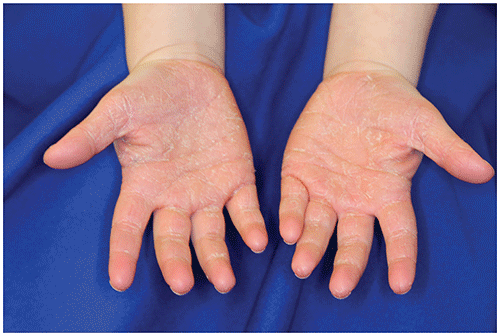
Figure 53.2. Plantar Involvement in Pityriasis Rubra Pilaris. Well-Demarcated Thickening of the Soles with Mild Scaling and Erythema in a Child with Juvenile Circumscribed Pityriasis Rubra Pilaris.
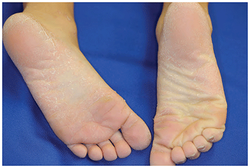
Figure 53.3. Well-Demarcated Erythematous Scaling Plaques with Follicular Prominence in a Young Child with Pityriasis Rubra Pilaris.
Look-alikes
| Disorder | Differentiating Features |
|---|
| Psoriasis | Characteristic body sites often involved (eg, scalp, postauricular skin, genitalia, extensor surfaces, periumbilical skin), although there is some overlap, as pityriasis rubra pilaris may involve similar areas. May have associated joint pain, swelling (ie, psoriatic arthritis). Thick, adherent silvery scales are characteristic.
|
| Pityriasis rosea | Acute onset of thin scaly plaques, often in a Christmas tree configuration on trunk. Larger plaque (herald patch) precedes development of smaller plaques. Lacks palmar and plantar involvement.
|
| Atopic dermatitis | Pruritic, scaly papules and plaques. Personal or family history of atopic diseases common. Characteristic body sites involved (eg, flexural surfaces in children; extensor involvement typically limited to infants). Typically lacks palmar and plantar involvement.
|
▶Typically diagnosed based on clinical features. In patients with atypical features or when the diagnosis is in question, biopsy may be warranted.
▶If diagnosis is in question, refer to a dermatologist.
▶Mild to moderate disease often responds to emollients, low- to mid-potency topical corticosteroids, or topical retinoids.
▶Keratolytics may help thin the hyperkeratosis on palms and soles.
▶Topical calcineurin inhibitors (eg, pimecrolimus, tacrolimus) may be helpful for facial involvement.
▶Severe disease is typically treated with systemic retinoids and, occasionally, immunosuppressants, such as methotrexate, cyclosporine, or azathioprine. Phototherapy has occasionally been helpful, and biologic therapies (namely, ustekinumab) have been reported to be useful in patients with CARD14 pathogenetic variants.
▶Prognosis is variable and difficult to predict and may be related to disease subtype. Remission is noted in a subset of children within a few months of onset, whereas others may have disease that persists for years.
▶Patients presenting with 1 form of PRP may occasionally have the PRP evolve into a different subtype.
▶If diagnosis is uncertain.
▶If patients have extensive disease or topical therapies fail.