▶Juvenile xanthogranulomas (JXGs) are benign nodular lesions occurring particularly in infants and young children.
▶They are collections of xanthomatous cells, but no association with systemic hyperlipidemia exists.
▶Lesions may be present at birth.
▶JXG is a common form of non-Langerhans cell histiocytosis; the course is self-limited, with resolution of lesions over several years.
▶Iris lesions can mimic retinoblastomas and may result in hyphema and/or glaucoma.
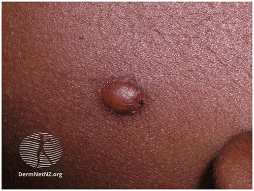
▶Characterized by orange or yellow-brown firm papules or papulonodular lesions (Figures 81.1 and 81.2). In patients with darker skin tones, they may be skin-colored or dark red or brown (Figure 81.3).
▶Early lesions erythematous; eventually become lipidized, with yellow color predominating clinically.
▶Often located in the head and neck area, although can be on any area of the body.
▶May be solitary or multiple.
▶Extra-cutaneous sites of involvement include eye (most common); less commonly, soft tissues, muscle, lung, liver, spleen, central nervous system, kidneys, and adrenal glands.
Figure 81.1. Juvenile Xanthogranuloma of the Scalp.
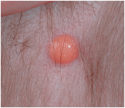
Figure 81.2. Juvenile Xanthogranuloma of the Scalp. A Small Yellow-Orange Papule.
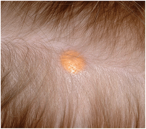
Figure 81.3. In Persons with Darker Skin Tones, a Juvenile Xanthogranuloma May be Skin Colored or, as in This Patient, Red-Brown. Reproduced with Permission from Dermnet Nz.
Look-alikes
| Disorder | Differentiating Features |
|---|
| Spitz nevus | Most often an erythematous papule. Does not undergo lipidization, so does not become yellow. May see brown pigment with dermoscopy or diascopy (compressing lesion and viewing through a glass slide).
|
| Solitary mastocytoma | |
| Melanocytic nevus | |
▶The diagnosis is usually made based on clinical findings.
▶Biopsy of the lesion will show foamy, multinucleated histiocytic giant cells with scattered eosinophils.
▶Observation and reassurance.
▶Most lesions resolve spontaneously over several years.
▶Surgical excision, when requested or clinically indicated (ie, for rapid growth, ulceration, or concern for cosmetic deformity).
▶Children with multiple or facial lesions should be referred to ophthalmology for eye examination.
▶Patients with neurofibromatosis type 1 and JXGs may have an increased risk of juvenile chronic myelogenous leukemia and should be monitored appropriately.
▶See the
Treating Associated Conditions
section.▶Consider referral to a dermatologist when the diagnosis is in question.
▶Neonates or infants with multiple lesions merit an evaluation for systemic involvement.