▶A multisystem autoimmune disorder with protean skin manifestations resulting from immune complex deposition and end-organ damage.
▶More common in female than male patients; approximately 80% of children and adults with systemic lupus erythematosus (SLE) are female; however, in prepubescent children, the male-to-female ratio may be more equal.
▶Presents most often in postpubertal female patients (20% of cases are diagnosed in the first 2 decades after birth), especially African-American, Asian, Hispanic, and Native American patients.
▶Median age of onset in childhood SLE is 11 to 12 years.
▶Exact cause remains poorly understood; believed to be related to genetic and environmental factors; hormonal factors may also play a role.
▶Extra-cutaneous targets most commonly include joints, hematologic system, lungs, heart, kidneys, and central nervous system.
▶Approximately 80% of patients with SLE will have skin involvement at some point in their course and often as the presenting feature.
▶Advances in early diagnosis and treatment have improved survival and quality of life for individuals who are affected; nevertheless, SLE can lead to significant morbidity and even mortality.
▶Malar erythema often seen; redness occurs in a butterfly distribution over the cheeks, sparing the nasolabial folds, and often appears after sun exposure.
■Edema often present along with facial erythema.
■Occasionally, rash has a papular component.
■Malar skin eruption usually transient and non-scarring.
▶Erythematous patches and papules over the dorsal aspect of the fingers may occur and usually spare the areas overlying joints (in contrast with juvenile dermatomyositis).
▶Discoid lupus erythematosus (DLE) lesions are a cutaneous manifestation seen in approximately 10% of children with SLE; DLE lesions are seen more commonly in adult SLE.
■Lesions usually are located on face (Figure 120.1), on or around ears, or on the scalp and are usually round or coin shaped, annular (central clearing present), hyperpigmented, and often scaly (Figures 120.2 and 120.3).
■Central atrophy may be present.
■Lesions vary in size, usually 1 to 3 cm.
■Often, lesions resolve with chronic pigmentary change (hypopigmentation or hyperpigmentation) and scarring (Figure 120.4).
■Approximately 25% to 30% of children with DLE will eventually progress to SLE, with the greatest risk being within the first year after the DLE diagnosis.
▶Non-scarring alopecia (hair loss) is commonly seen in SLE but is nonspecific; it most often presents as thinning of the hair in the temporal scalp regions.
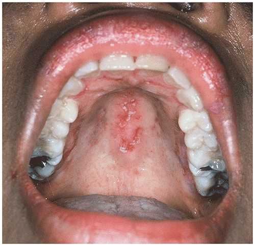
▶Nasal, oral, and palatal ulcerations (Figure 120.5); dilated nail fold capillaries (telangiectasias); petechial or purpuric lesions; livedo reticularis; erythema nodosum; photosensitivity; and small ice pick–like scars of the fingertips are other cutaneous features of SLE.
Figure 120.1. Teenage Patient with Systemic Lupus Erythematosus and Facial Lesions of Discoid Lupus Erythematosus with Erythema, Atrophy, and Hyperpigmentation.
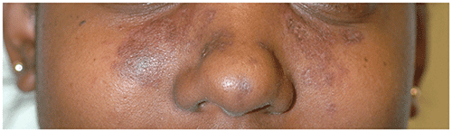
Figure 120.2. Lesions of Active Discoid Lupus Erythematosus on the Arm in Addition to Areas of Postinflammatory Hyperpigmentation and Scarring in Sites of Previous Lesions.
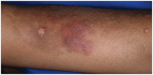
Figure 120.3. (A) Multiple Erythematous Papules and Plaques on the Face of a Boy with Chronic Cutaneous (Discoid) Lupus Erythematosus. Note the Cutaneous Atrophy of Several Lesions, Particularly on the Earlobe, a Characteristic Location for Lupus Lesions. (B) Erythematous Papules on the Cheek of the Same Patient with Discoid Lupus Erythematosus Shown in Figure 120.3a.
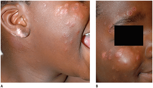
Figure 120.4. Atrophic Crusted Ulcerations on the Face of an Adolescent Who Has Discoid and Systemic Lupus Erythematosus.
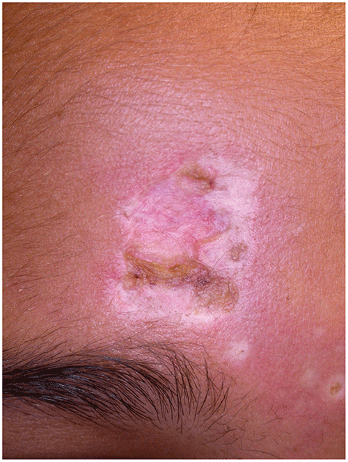
Figure 120.5. Palatal Ulcerations in This Young Adult with Systemic Lupus Erythematosus.
Other Clinical Findings
▶Fever, malaise, weight loss, and arthralgias or arthritis are common in children with SLE.
▶Additional signs and symptoms include fatigue, abdominal pain, muscle weakness, lymphadenopathy, hepatosplenomegaly, anorexia, weight loss, night sweats, and Raynaud phenomenon (blanching of fingertips with cold exposure followed by cyanosis and a reactive hyperemia on rewarming).
▶Pulmonary (most often pleuritis) and cardiac (including pericarditis, myocarditis, valvular disease, and coronary artery vasculitis) manifestations may also occur in children with SLE.
Lupus Variants
Discoid Lupus Erythematosus (DLE)
▶Discoid lesions may be seen in the setting of SLE or in patients with skin disease only.
▶DLE is also known as chronic cutaneous lupus erythematosus.
▶Skin lesions present as annular, scaly plaques with pigmentary change and atrophy (see Figures 120.1–120.4). Early discoid lesions may occasionally be confused with tinea corporis (ringworm).
▶Scarring may occur.
▶Lesions of DLE are often exacerbated by sun exposure.
Subacute Cutaneous Lupus Erythematosus (SCLE)
▶Subacute cutaneous lupus erythematosus (SCLE) is a subtype of lupus characterized by significant photosensitivity; it only occasionally occurs in children.
▶Lesions usually appear in a sun-exposed distribution, and, in most, the condition is milder in severity than SLE.
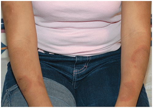
▶Lesions are often annular or psoriasis-like in configuration (Figure 120.6).
▶Postinflammatory pigmentary changes are common, but scarring does not occur.
▶Most patients have positive anti–SS-A (anti-Ro) antibody, which is associated with photosensitivity; pregnant women with anti–SS-A antibody (whether or not they have overt SLE or SCLE) are at risk of having a neonate with neonatal lupus erythematosus (NLE); anti–SS-B (anti-La) antibody is also associated with SCLE.
▶Approximately 15% of patients with SCLE develop significant systemic disease with time; in general, though, patients with SCLE may have a better prognosis than those who have SLE.
Figure 120.6. Large Edematous, Erythematous, Arcuate, and Annular Plaques on the Arms of This Teenager with Subacute Cutaneous Lupus Erythematosus.
Neonatal Lupus Erythematousus (NLE)
▶A distinct type of lupus seen in newborns and young infants that results from transplacentally acquired autoantibodies, most often anti–SS-A (anti-Ro) antibody or anti–SS-B (anti-La) antibody, potentially in association with congenital heart block.
▶Antibodies to U1 ribonucleoprotein (RNP) may also be associated with NLE and are usually not associated with congenital heart block.
▶Most common target organs in NLE are the skin and heart.
▶Approximately one-half of patients with NLE have cutaneous manifestations.
▶NLE is the most common cause of congenital heart block; unfortunately, it often results in third-degree heart block requiring pacemaker placement.
▶Skin lesions usually develop between birth and 8 weeks of age and are distributed most commonly on the face and head; parents often report exacerbation or appearance after sun exposure.
▶Skin lesions are usually annular and erythematous or telangiectatic; they are often mildly scaly and may resemble lesions of tinea corporis (Figure 120.7), seborrheic dermatitis, or atopic dermatitis. Mild atrophy may be present.
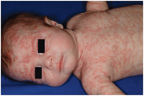
▶Although some infants with NLE have lesions in photodistribution (in areas of sun exposure), others may have more generalized eruptions involving skin that was never exposed to the sun (Figure 120.8).
▶There may be a distinct periorbital accentuation, termed the “raccoon eyes” appearance.
▶Lesions of NLE are self-limited and usually resolve by 6 months of age without scarring.
▶Approximately 10% of infants may develop hepatic or hematologic alterations (usually self-limited).
▶Fewer than one-half of mothers have a known diagnosis of autoimmune disease; mothers are most likely to have (or eventually develop) SCLE, SLE, or Sjögren syndrome.
▶Infants with cutaneous findings suggestive of NLE should be evaluated for cardiac, hematologic, or hepatic abnormalities; serological studies usually confirm the diagnosis and should be performed in infant and mother.
▶Skin biopsy is rarely indicated.
▶Mothers who do not have symptoms but have infants with NLE should undergo comprehensive rheumatologic evaluation and be followed up closely for connective tissue disease.
▶There is an increased risk of having another infant with NLE in subsequent pregnancies.
Figure 120.7. Multiple Annular Plaques with Dusky Atrophic Centers on the Face and Scalp of This 1-Month-Old with Neonatal Lupus Erythematosus. The Mother Had No Previous History of Connective Tissue Disease but Later Received a Diagnosis of Systemic Lupus Erythematosus. Her Subsequent Pregnancy Resulted in a Second Child with Cutaneous Neonatal Lupus Erythematosus.
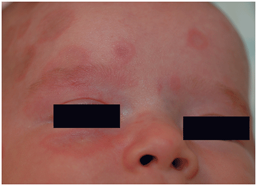
Figure 120.8. Young Infant with Widespread Erythematous, Slightly Atrophic Patches and Plaques Secondary to Neonatal Lupus Erythematosus. Note the Prominent Involvement of the Periorbital Region, Forehead, and Scalp.
Look-alikes
| Disorder | Differentiating Features |
|---|
| Systemic Lupus Erythematosus |
|---|
| Juvenile idiopathic arthritis, systemic subtype | Evanescent hive-like lesions. Usually no malar erythema. High-spiking fevers common, often correlate with the presence of skin eruption. Skin findings may exhibit Koebner phenomenon.
|
| Juvenile dermatomyositis | Proximal muscle weakness in most patients; less common in systemic lupus erythematosus. Arthritis usually absent. Distribution of cutaneous lesions over extensor surfaces of arms and legs. Heliotrope rash (periorbital distribution). May have malar rash, which often does not spare nasolabial folds. Skin changes often show lilac color. Calcinosis cutis can occur in some patients. Gottron sign (erythema) or papules (lichenoid papules) present over the knuckles and occasionally elbows, knees, and ankles.
|
| Drug hypersensitivity syndrome (sulfa, minocycline, aromatic anticonvulsants, lamotrigine) (also known as drug reaction with eosinophilia and systemic symptoms [DRESS]) | Usually classic triad of fever, rash, and lymphadenopathy. History of drug ingestion, usually preceding syndrome by 3 to 8 weeks. Increased eosinophils and atypical lymphocytes on complete blood cell count. Histologic findings distinct from those of lupus. Sudden onset of diffuse cutaneous eruption; distinct facial or periorbital edema often present. Eventual exfoliation; some patients with generalized exfoliative erythroderma. Frequent liver involvement; occasionally fulminant hepatitis.
|
| Rosacea | No systemic abnormalities. Often more prominent in the nasolabial folds and over medial aspect of the cheeks. Inflammatory papules and pustules may be present. Facial telangiectasias common. May involve the eyes (ocular rosacea). Chronic waxing and waning course. Exacerbated by sunlight, heat, alcohol, hot beverages, spicy foods.
|
| Polymorphous light eruption | Lacks systemic associations. Typically presents in late spring with initial sun exposure of the season. Edema and erythema of the face, ears, arms, and dorsal aspect of the hands.
|
| Subacute Cutaneous Lupus Erythematosus |
|---|
| Psoriasis | Most often occurs on knees, elbows, scalp, umbilicus, gluteal crease. Lesions are plaques, typically thicker than subacute cutaneous lupus erythematosus lesions, and have silvery to white (micaceous) adherent scale. May have associated nail findings. Typically improves (rather than worsens) in sunlight. Negative serological study results for lupus erythematosus.
|
| Tinea corporis | Plaques usually have more scaling and may have central clearing. Inflammatory papules or pustules may be present. No accentuation in sun-exposed sites. Positive potassium hydroxide preparation or fungal culture results. Negative serological study results for lupus erythematosus.
|
| Granuloma annulare | Annular smooth plaques that lack scale and are not exacerbated by sunlight. Most often distributed on dorsal aspect of feet, ankles, wrists, and legs. No systemic abnormalities in most patients. Negative serological study results for lupus erythematosus. Resolves spontaneously over 2 to 4 years.
|
| Neonatal Lupus Erythematosus (None of the following conditions are associated with increased risk of
congenital heart block, and in each of them, serological
study results for neonatal lupus erythematosus would be negative.) |
|---|
| Seborrheic dermatitis | May also have scalp dermatitis, usually with greasy, adherent yellow scaling. Erythema and maceration may occur in the neck, inguinal and axillary folds, and umbilicus (and, less often, the popliteal and antecubital fossae). Onset in early infancy. Usually self-limited with spontaneous clearing by 6 to 12 months of age.
|
| Tinea corporis or faciei | Positive potassium hydroxide preparation or fungal culture results. May have more scale or inflammatory papules or pustules. No accentuation in sun-exposed areas. No periorbital accentuation. History of known exposure may be present.
|
| Psoriasis | Facial involvement less common. Often shows significant diaper and umbilical involvement in infant. No accentuation in sun-exposed sites. Typically improves (rather than worsening) with sun exposure.
|
| Atopic dermatitis | Pruritus very common. No typical accentuation in sun-exposed areas. Associated excoriations or crusting may be present. Lichenification (thickening of skin in chronically rubbed sites) often present. Lesions not typically annular. Atopic diathesis often present.
|
▶A thorough history, review of systems, and physical examination are critical in the accurate diagnosis of lupus. Helpful physical findings in SLE include the following:
■Malar erythema
■Nasal or oral ulcerations
■Diffuse non-scarring alopecia
■Raynaud phenomenon
■Periungual telangiectasia
■Vasculitis (red or purple macules and papules on hands or small ulcerations of the fingertips)
■Lymphadenopathy
■Erythema of the palms
▶When a diagnosis of SLE is suspected, the following laboratory evaluations should be considered:
■Antinuclear antibody profile (including anti-dsDNA, anti-Sm, anti–SS-A [anti-Ro], anti–SS-B [anti-La], anti-RNP antibodies)
■Antiphospholipid antibody and lupus anticoagulant panels
■Complete blood cell count with differential and platelet counts
■Chemistries to include liver and renal function
■Complement levels (C3, C4, total hemolytic complement)
■Erythrocyte sedimentation rate (less commonly, C-reactive protein level)
■Urinalysis with microscopic examination and first-morning spot ratio of protein to creatinine
▶Serological studies may aid in confirming the diagnosis of lupus and may also help with categorization of subtype and prognosis.
■Antinuclear antibody almost always positive in SLE but is also positive in 5% to 10% of the general population.
■If high clinical suspicion of lupus, check for anti–native dsDNA antibodies; these are highly specific for SLE and present in approximately one-half of patients; often associated with renal disease.
■Antibodies against small nuclear RNPs.
– Anti-Smith: specific for SLE; present in 20% of patients with SLE; associated with higher risk for renal disease.
– Anti-RNP: may be seen in SLE, scleroderma, NLE, or mixed connective tissue disease.
– Anti–SS-A (anti-Ro): seen in approximately 30% of patients with SLE and approximately one-half of patients with Sjögren syndrome, as well as in patients with SCLE and NLE; strong association with photosensitivity.
– Anti–SS-B (anti-La): positive in approximately 10% patients with SLE; often seen in association with anti–SS-A (anti-Ro) antibody; also seen in SCLE and NLE.
■Antiphospholipid antibody: occurs in 30% to 50% of adult patients with lupus and can also occur in patients with antiphospholipid antibody syndrome; associated with higher incidence of thrombotic events; skin findings may include livedo reticularis or cutaneous ulcerations.
■Patients with SLE should meet the new 2019 European League Against Rheumatism/American College of Rheumatology classification criteria for SLE according to the following weighted scoring system:
Table 120.1. The 2019 European League Against Rheumatism/American College of Rheumatology Classification Criteria
| Additive Criteria |
|---|
| Clinical domains and criteria | Weight |
|---|
| Constitutional | |
|---|
| Fever | 2 |
| Hematologic | |
|---|
| Leukopenia | 3 |
| Thrombocytopenia | 4 |
| Autoimmune hemolysis | 4 |
| Neuropsychiatric | |
|---|
| Delirium | 2 |
| Psychosis | 3 |
| Seizure | 5 |
| Mucocutaneous | |
|---|
| Non-scarring alopecia | 2 |
| Oral ulcers | 2 |
| Subacute lupus OR discoid lupus | 4 |
| Acute cutaneous lupus | 6 |
| Serosal | |
|---|
| Pleural or pericardial effusion | 5 |
| Acute pericarditis | 6 |
| Musculoskeletal | |
|---|
| Joint involvement | 6 |
| Renal | |
|---|
| Proteinuria >0.5 g/24 h | 4 |
| Renal biopsy Class II or V lupus nephritis | 8 |
| Renal biopsy Class III or IV lupus nephritis | 10 |
| Immunology domains and criteria | Weight |
|---|
| Antiphospholipid antibodies | |
|---|
| Anticardiolipin antibodies OR
Anti-β2GP1 antibodies OR
Lupus anticoagulant | 2 |
| Complement proteins | |
|---|
| Low C3 OR low C4 | 3 |
| Low C3 AND low C4 | 4 |
| Systemic lupus erythematosus–specific antibodies | |
|---|
| Anti-dsDNA antibody OR
Anti-Smith antibody | 6 |
Systemic lupus erythematosus classification requires antinuclear antibodies at a titer of 1:80 or greater, at least 1 clinical criterion, and >10 points. Adapted from Aringer M, Costenbader K, Daikh D, et al. 2019 European League Against Rheumatism/American College of Rheumatology classification criteria for systemic lupus erythematosus. Arthritis Rheumatol. 2019;71(9):1400–1412.
■Traditional clinical and laboratory criteria that were used to diagnose SLE are listed here (American College of Rheumatology 1997 revised criteria for classification of SLE); SLE criteria are met if the patient has at least 4 of the 11 following:
Malar rash
Discoid rash
Photosensitivity
Oral ulcerations
Oral or nasopharyngeal ulcers, usually painless
Arthritis
Two or more joints
Nonerosive arthritis
Serositis (pleuritis or pericarditis)
Renal disease (persistent proteinuria or cellular casts)
Neurologic manifestations (seizures, psychosis)
Hematologic disorder (≥1 of the following findings on >1 occasion):
Hemolytic anemia
Leukopenia (<4,000/mm3)
Lymphopenia (<1,500/mm3)
Thrombocytopenia (<100,000/mm3)
Immunologic disorder (≥1 of the following):
Anti-DNA antibody to native DNA in abnormal titer.
Anti-Sm antibody.
Positive finding of antiphospholipid antibodies on the basis of an abnormal serum level of immunoglobulin G or immunoglobulin M anticardiolipin antibodies, a positive test result for lupus anticoagulant according to a standard method, or a false-positive serological test result for syphilis known to be positive for at least 6 months and confirmed with Treponema pallidum immobilization or fluorescent treponemal antibody absorption test.
Antinuclear antibody (in absence of drugs known to cause drug-induced lupus).
▶Patients who have SLE should be followed up closely by a pediatric rheumatologist, and a multidisciplinary approach should be used for those who have significant systemic disease.
▶Cutaneous manifestations may be managed in conjunction with a pediatric or adult dermatologist.
▶Treatment of cutaneous disease may include (depending on severity and response to treatment) the following:
■Photoprotection
– Avoidance of excessive sun exposure, especially between 10:00 am and 4:00 pm.
– Sun protective clothing (clothing with ultraviolet protection factor 50 rating, wide-brimmed hats, ultraviolet-protective sunglasses).
– Daily broad-spectrum sunscreen use with high sun protection factor (≥30); products containing titanium dioxide or zinc oxide are optimal.
■Topical or intralesional corticosteroids
■Topical calcineurin inhibitors (tacrolimus, pimecrolimus)
■Systemic agents for more severe skin disease
▶Treatment of systemic disease usually requires one or a combination of the following systemic medications:
■Corticosteroids
■Azathioprine
■Mycophenolate mofetil
■Methotrexate
■Cyclophosphamide
■Rituximab
■Belimumab, an anti–B-lymphocyte stimulator antibody
■Intravenous immunoglobulin
▶In addition, nonsteroidal anti-inflammatory agents can be used for musculoskeletal symptoms and serositis but should not be used in patients with nephritis.
▶Patients with SLE are likely to need concurrent care from multiple specialists depending on the degree of end-organ involvement.
▶Those with significant renal disease often benefit from collaborative care with a nephrologist.
▶Those with neuropsychiatric disease may require specialty care directed at central nervous system complications.
▶Because of the chronic and potentially disabling nature of lupus, the disorder can be associated with significant psychologic morbidity, and care from an appropriate specialist may be very beneficial.
▶The prognosis for patients with SLE is quite variable and depends on the organ systems involved.
▶Patients who have diffuse proliferative glomerulonephritis and associated hypertension often have a poorer prognosis.
▶Infection is a major cause of death and is usually related to use of systemic corticosteroids or other immunosuppressive agents.
▶Children with DLE generally have a good prognosis; however, these patients may go on to develop systemic disease (SLE) and should be followed up clinically and with intermittent laboratory evaluations.
▶Infants with NLE have a good overall prognosis; those with congenital heart block often require pacemaker placement.
▶Children suspected of having cutaneous or systemic manifestations of lupus should be referred to a rheumatologist or dermatologist for confirmation of diagnosis and management.
▶If a patient with SLE acutely develops cytopenia and fever, the diagnosis of macrophage activation syndrome should be considered and is often accompanied by hyperferritinemia and hypertriglyceridemia; these patients should immediately be evaluated by a pediatric rheumatologist.
▶Infants suspected of having NLE should undergo immediate cardiac evaluation, including electrocardiography, to assess for congenital heart block; mothers of these infants should be referred for rheumatologic evaluation.
▶Lupus Foundation of America: Provides information, support, and links for patients and families.
www.lupus.org
▶Lupus Initiative: Provides information and support for patients, families, and medical professionals. Sponsored by the American College of Rheumatology.
www.thelupusinitiative.org
▶Lupus Research Alliance: Provides information and support for patients and families.
https://www.lupusresearch.org