▶Dermatophyte infection of the feet; organisms responsible are Trichophyton rubrum, Trichophyton mentagrophytes, and Epidermophyton floccosum.
▶Common in adolescents and adults; less common in childhood.
▶Warm, moist environment of occlusive footwear predisposes to fungal infection.
Three forms of infection are recognized: interdigital, vesicular, and moccasin.
▶Interdigital
■Typically caused by T rubrum or E floccosum.
■Pruritus, erythema, fissuring, scaling, and maceration occur in the interdigital spaces (Figure 40.1).
▶Vesicular
■Typically caused by T mentagrophytes.
■Vesicles, bullae, and erosions appear on the instep of the foot or dorsal aspect of the foot or between the toes (Figure 40.2).
▶Moccasin
■Typically caused by T rubrum or E floccosum.
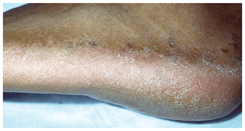
■Erythema and scaling involve much of or all the plantar surface and sides of the feet (Figure 40.3).
▶Rarely, a dermatophytid (id) or autosensitization reaction occurs that produces a widespread eczematous-appearing eruption composed of papules or deep-seated vesicles.
Figure 40.1. Erythema, Scaling, Fissuring, and Erosions Between the Toes are Seen in the Interdigital Form of Tinea Pedis.
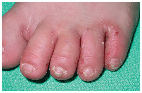
Figure 40.2. Erythematous Plaques, Maceration, and Toe Web Bullae in a Child with Vesicular Tinea Pedis.
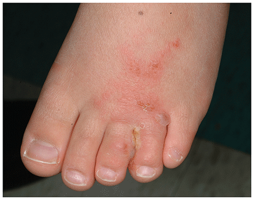
Figure 40.3. Moccasin Form of Tinea Pedis with Erythema and Scaling Involving the Plantar Surface and Side of the Left Foot. Reproduced with Permission from Ely Jw, Rosenfeld S, Seabury Stone M. Diagnosis and Management of Tinea Infections. Am Fam Physician. 2014;90(10):702–710.
Look-alikes
In each of the conditions listed herein, a potassium hydroxide preparation or fungal culture would fail to confirm the presence of fungal infection.
| Disorder | Differentiating Features |
|---|
| Contact dermatitis | |
| Erythrasma | Often red-brown or brown; involves interdigital spaces. Elevated border and scaling absent. Wood lamp examination reveals coral red fluorescence.
|
| Juvenile plantar dermatosis | Intense erythema with fissuring on the plantar surface of the foot, most typically the ball and heel, often with sparing of arch. History of hyperhidrosis common. Interdigital spaces spared. History of atopic dermatitis often present.
|
| Pitted keratolysis | Small pits that may coalesce into larger, very superficial erosions present on plantar surface of foot. Hyperhidrosis and malodor often present. Interdigital spaces spared.
|
▶The diagnosis is made clinically.
▶If uncertainty exists, a potassium hydroxide preparation (revealing branching hyphae) or fungal culture (eg, dermatophyte test medium) may be performed (see Figure 37.5).
▶For typical infections, application of a topical antifungal agent, such as an imidazole (eg, clotrimazole, miconazole, ketoconazole), allylamine (eg, terbinafine, naftifine), or tolnaftate, is appropriate. The agent is applied until the eruption clears, typically within 3 to 4 weeks.
▶Widespread, resistant, or severe infections may require oral therapy with griseofulvin or another antifungal agent.
▶Advise patients to keep their feet dry and, if possible, to wear well-ventilated shoes or sandals.
▶For patients who experience recurrences, recommend the regular use of an absorbent powder containing an antifungal agent (eg, Zeasorb AF, Micatin, Tinactin, Desenex, Lotrimin AF, and others).
▶If treatment of concomitant nail infection (ie, onychomycosis) is desired, oral therapy with terbinafine or itraconazole will be required.
▶The prognosis is excellent.
▶To prevent recurrences, advise patients to dry carefully after bathing or showering, wear protective footwear in public showers, wear well-ventilated shoes or sandals, and regularly apply an absorbent powder containing an antifungal agent (eg, Zeasorb AF, Micatin, Tinactin, Desenex, Lotrimin AF, and others).
▶Consider consultation with a dermatologist when the diagnosis is in doubt or when appropriate therapy fails.